Almost every animal cell diagram in circulation gives the nucleus about a third of the picture and draws two mitochondria beside it. The measured numbers for a liver cell are the other way round: the nucleus is 6% of the volume and mitochondria are 22%. That one inversion is the reason a generation of students believes the nucleus is the biggest thing in a cell, and it is fixable in the drawing.
Quick answer
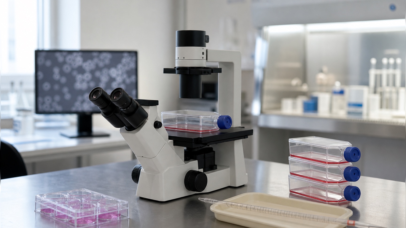
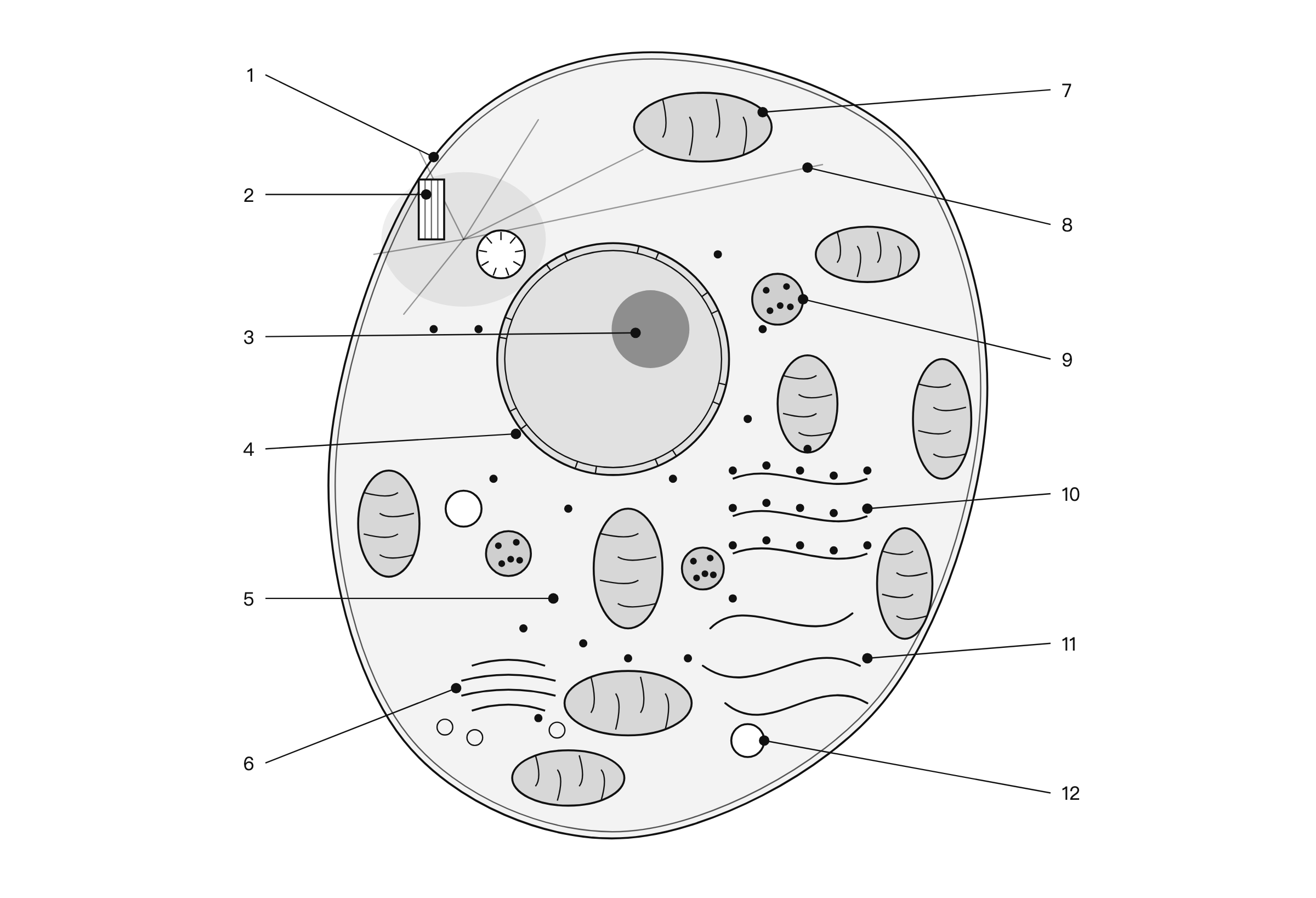
An animal cell diagram is a labelled section through one cell. Working outward from the centre: nucleolus inside the nucleus, whose double envelope is pierced by nuclear pores; rough and smooth endoplasmic reticulum, which are one membrane system; Golgi apparatus; mitochondria; lysosomes and peroxisomes; the centrosome with its pair of centrioles; ribosomes and the cytoskeleton in the cytosol; and the plasma membrane, which is the outermost boundary because there is no wall outside it.
What does an animal cell diagram show?
It shows a section. An animal cell is a three-dimensional object and the diagram cuts it open, which means the outline you are looking at is a cut edge, not a silhouette.
Three things on the diagram are claims. Which compartments are present. How they nest inside one another. And which membranes are continuous with which — the part almost every diagram draws and almost no reader is told to look for. The rough and smooth ER are one system. The nuclear envelope is continuous with the ER. The plasma membrane is continuous with nothing; it is the edge.
Everything else is a drawing decision. The colours are conventional, not observed. The number of each organelle drawn is whatever fitted. And the sizes are wrong in a specific, correctable direction, which the next two sections are about.
One thing that is not a drawing decision: the shape. A plant cell is drawn as a box because a wall holds it in one. An animal cell has no wall, so the outline is the plasma membrane and nothing else, and it takes whatever shape the cytoskeleton and its neighbours give it. Drawing an animal cell as a neat circle is the first small lie in most diagrams.
The parts of an animal cell, labelled
Each label, and what the structure behind it actually is:
| Labelled part | What it is | What it does |
|---|---|---|
| Plasma membrane | A lipid bilayer, and the whole of the cell’s outer boundary. Only about 2% of the total membrane in a liver cell is plasma membrane | Controls what enters and leaves. In an animal cell it also does the structural job a wall does in a plant, which is why the cell has no fixed shape |
| Cytosol | The aqueous phase the organelles are suspended in — a little more than half the total volume of the cell | Protein synthesis and protein degradation. Held near pH 7.2, which is why a leaking lysosome does little damage |
| Nucleus | A double-membraned envelope enclosing the chromosomes, pierced by nuclear pores, with a nucleolus inside. About 6% of a liver cell’s volume | Holds the genome; the principal site of DNA and RNA synthesis |
| Nucleolus | A dense, unbounded region inside the nucleus — no membrane of its own | Ribosome subunit assembly. It is drawn solid because it is denser than the surrounding nucleoplasm, not because it is enclosed |
| Rough endoplasmic reticulum | Flattened cisternae studded with ribosomes on the cytosolic face. Its cisternae are 9% of cell volume; its membrane is 35% of all the membrane in a liver cell and 60% in a pancreatic exocrine cell | Synthesis and folding of proteins entering the secretory pathway |
| Smooth endoplasmic reticulum | The same continuous membrane system, tubular rather than flattened, with no ribosomes bound to it | Lipid synthesis, detoxification, calcium storage. It is not a separate organelle — the difference from rough ER is what is on the surface |
| Golgi apparatus | A stack of flattened cisternae with vesicles budding from its faces, usually close to the nucleus. Its cisternae plus the smooth ER cisternae come to 6% of cell volume | Modifies, sorts and dispatches proteins from the ER; adds the mannose 6-phosphate tag that routes hydrolases to lysosomes |
| Mitochondrion | A double-membraned organelle 0.5–1 μm across whose inner membrane is folded into cristae. Together they are 22% of a liver cell’s volume, and the inner membrane alone is 32% of all its membrane | Respiration-driven ATP synthesis. The cristae are what the folding buys: surface area for the machinery |
| Lysosome | A single-membraned compartment holding acid hydrolases, kept at about pH 5.0 by an ATP-driven proton pump. About 1% of cell volume | Degradation of macromolecules delivered by endocytosis and autophagy. The enzymes only work at acid pH, which is the safety mechanism |
| Peroxisome | A small compartment with a single membrane, containing neither DNA nor ribosomes. About 1% of cell volume | Fatty acid oxidation, plasmalogen synthesis, and other oxidative reactions. Its proteins are made on free cytosolic ribosomes and imported fully folded |
| Centrosome | A pair of centrioles at right angles to each other, each a cylinder of nine triplets of microtubules, surrounded by amorphous pericentriolar material | The major microtubule-organising centre of an animal cell. It duplicates in interphase and gives rise to the two poles of the mitotic spindle |
| Ribosomes | Free in the cytosol, bound to the rough ER, and present again inside mitochondria | Translation. The separate set inside mitochondria is the visible remnant of their bacterial ancestry |
| Cytoskeleton | Three filament systems: actin filaments about 7 nm across, intermediate filaments, and microtubules — rigid hollow rods about 25 nm across | Shape, mechanical strength, transport and movement. In an animal cell it does the job the wall does in a plant, and it is the reason the outline is irregular |
How much of an animal cell is each organelle?
This is the part almost no diagram gets right, and the numbers have been in print since 1983.
Here is the measured breakdown for a liver cell, from Alberts' Table 12-1. A hepatocyte is not every animal cell — a pancreatic exocrine cell is five times smaller and packs sixty per cent of its membrane into rough ER — but it is a real cell with real numbers, which is more than a diagram drawn from another diagram can say.
| Compartment | Percentage of total cell volume |
|---|---|
| Cytosol | 54 |
| Mitochondria | 22 |
| Rough ER cisternae | 9 |
| Smooth ER cisternae plus Golgi cisternae | 6 |
| Nucleus | 6 |
| Peroxisomes | 1 |
| Lysosomes | 1 |
| Endosomes | 1 |
Relative volumes occupied by the major intracellular compartments in a liver cell, from Alberts et al., Molecular Biology of the Cell, 4th edition, Table 12-1.
Two things fall out of that table.
Mitochondria occupy nearly four times as much of the cell as the nucleus. Not slightly more. Nearly four times. The standard diagram — big central nucleus, two mitochondria off to the side — inverts the relationship, and it is the single most consistent error in the genre.
The membrane-enclosed compartments together take about half the cell. The cytosol is the other half. A diagram that draws the organelles as small islands in a large empty interior has the proportion backwards; the interior is not empty, and there is no more of it than there is of everything else.
The figure above is drawn against the convention. Measured off the drawing, the nucleus covers 10.3% of the section and the nine mitochondria together cover 12.1% — so the mitochondria come out larger, which is the direction the table demands.
It is still an understatement, and the size of the understatement is worth stating. The true ratio is 22 to 6, or 3.7 to 1; the figure manages 1.2 to 1. Nine mitochondria drawn large enough to carry a leader line do not add up to 22% of the picture, and no labelled diagram can — the moment the organelles are small enough to be correctly proportioned, they are too small to label. That is a real limit of the format, and it is better said out loud than quietly drawn around.
What is the difference between a plant cell and an animal cell?
The differences that matter to a diagram are structural, and they run in both directions.
| Feature | Plant cell | Animal cell |
|---|---|---|
| Outer boundary | Plasma membrane inside a cellulose cell wall, at least 0.1 μm thick between neighbouring cells | Plasma membrane only. Nothing outside it |
| Shape | Held by the wall — drawn as a box, and it is one | Held by the cytoskeleton and by neighbouring cells. Irregular |
| Plastids | Present in all living plant cells: chloroplasts, chromoplasts, amyloplasts | Absent |
| Large central vacuole | Usually over 30% of cell volume, up to 90% in some cell types | Absent. Small vesicles and endosomes only |
| Lysosomes | The vacuole is the related compartment and carries the same hydrolases | Present as discrete organelles at about 1% of cell volume, held near pH 5.0 |
| Centrioles | Absent from higher plants | A pair inside the centrosome, each nine triplets of microtubules |
| Cell-to-cell channels | Plasmodesmata: cytoplasmic channels 20–40 nm across, crossing the wall | Gap junctions: channels with a pore about 1.5 nm across |
| Other junctions | None needed — the middle lamella cements the walls together | Tight junctions seal sheets; desmosomes anchor intermediate filaments across cells |
| Cell division | A new wall is built outward from inside the cell | The cell pinches itself in two with a contractile ring |
The reversal worth noticing is the last three rows. Plant cells need no anchoring junctions because their walls are already glued together, so the whole apparatus of tight junctions, desmosomes and gap junctions is an animal solution to a problem plants solved with cement. That is why a plant cell diagram and an animal cell diagram look most different where two cells touch — and why the next section draws that.
How two animal cells connect
A plant cell talks to its neighbour through a plasmodesma: a cytoplasmic channel 20–40 nm across that crosses a wall at least 0.1 μm thick, with the plasma membrane of one cell running continuously into the other.
An animal cell has no wall to cross and does it differently. At a gap junction the two plasma membranes come within 2–4 nm of each other, and matched half-channels called connexons dock across that gap to form a continuous pore from one cytoplasm to the other.
The 1.5 nm figure comes from injecting fluorescent molecules of known size into one cell and watching what arrives in the next. Below about 1000 daltons — inorganic ions, sugars, amino acids, nucleotides, vitamins, and second messengers like cyclic AMP — molecules cross freely. Above it, they do not, so coupled cells share their small molecules but keep their proteins and nucleic acids to themselves.
The other two junctions belong on a diagram of an epithelium rather than of a single cell, and they are worth naming because they are what the single-cell diagram leaves out. Tight junctions seal neighbouring cells together into a sheet, so that a tracer applied on one side does not pass beyond them, and they also fence the plasma membrane into an apical and a basolateral domain that do not mix. Desmosomes are button-like contacts that rivet cells together and anchor intermediate filaments from one cell to the next, forming a framework of great tensile strength across a whole tissue.
How do you draw an animal cell?
Six decisions, in the order they matter.
Draw the outline irregular. Not a circle, not a rounded box. Nothing outside the membrane holds the cell to a shape, and an irregular outline says so before a single label is read.
Put the nucleus in at about a tenth of the area. A sphere at 6% of a cell's volume cuts a central section around 15% of the cell's area, and a diagram that gives it a third is claiming something the numbers do not support. Draw it off-centre while you are at it — the centrosome sits beside it, and the Golgi is usually close to it.
Draw more mitochondria than feels right. Two is the convention and two is wrong. Enough of them that they collectively look larger than the nucleus is closer to the truth, and still short of it.
Draw the rough and smooth ER as one system. Cisternae with ribosomes on the cytosolic face running into bare tubules. Two separate blobs in different corners is the commonest way of teaching a fact that is not true.
Give each organelle one distinguishing feature. A mitochondrion without cristae and a lysosome are the same oval. The cristae are the distinction, so draw them. Nine ticks around an end-on centriole are the distinction between a centriole and a capsule, so draw those too.
Then label it. Leader lines straight, none crossing another, each ending in a dot on the structure it names. Set the type at a real size while you draw: Nature allows a maximum of 7 pt for figure text and a minimum of 5 pt, with 8 pt bold for panel letters, in a figure 89 mm wide for one column or 183 mm for two, on a page 247 mm deep. A cell diagram drawn at poster scale and dropped into a manuscript at a third of the size is the most common way a good figure becomes an unreadable one.
Vector formats survive that reduction and raster formats do not. If the diagram has to be raster, Nature wants a minimum of 300 dpi at the size it will be printed.
The same diagram without the labels
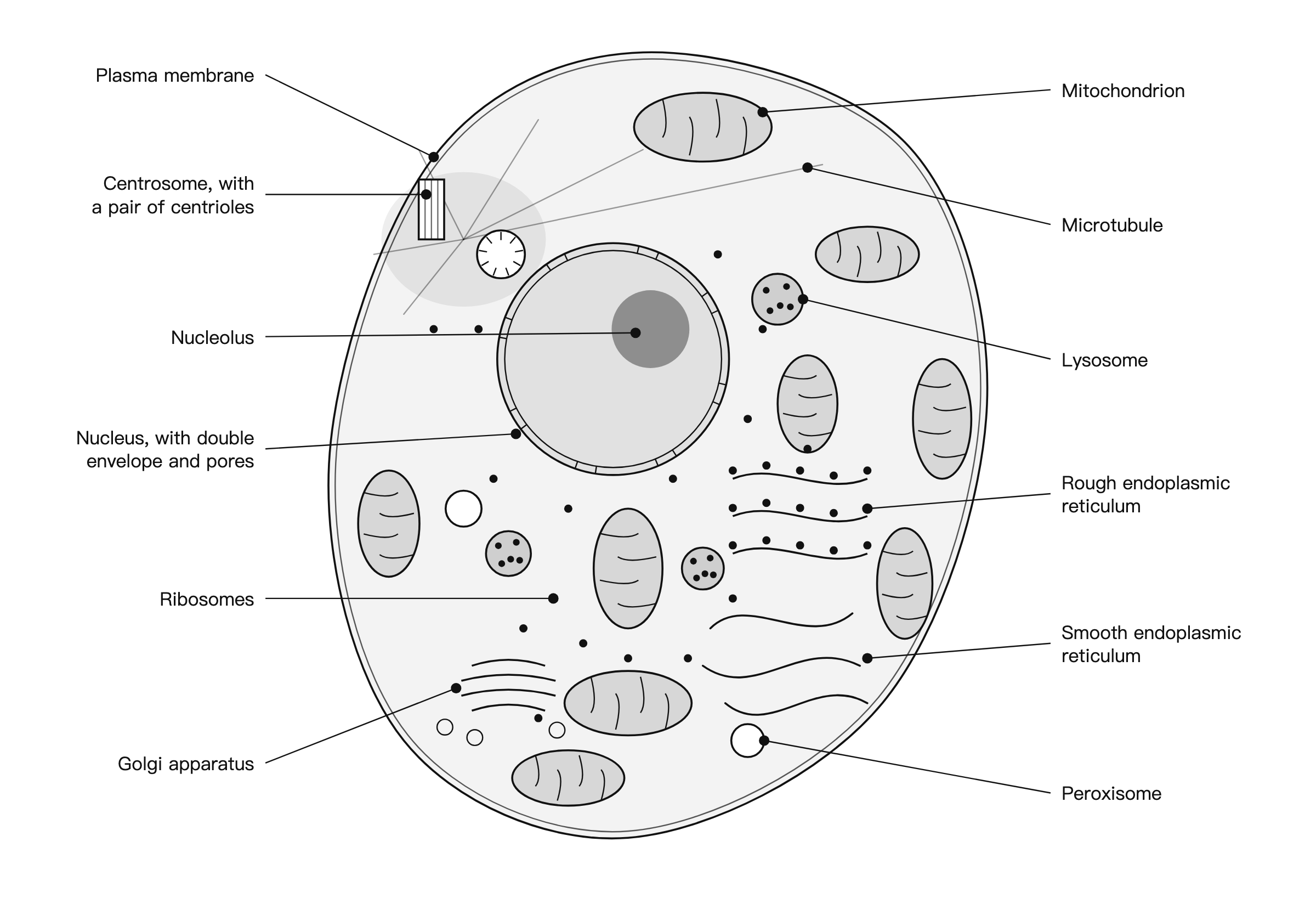
The same geometry with numbers instead of names. Print it, label it from memory, then check against the table above.
Five things animal cell diagrams get wrong
The nucleus is too big and the mitochondria too few. Covered above, and it is the one that changes what a reader believes about the cell.
Rough and smooth ER drawn as two organelles. They are one continuous membrane system. The only difference is whether ribosomes are bound to the cytosolic face, and a diagram that puts them in opposite corners with separate labels teaches the opposite.
The nucleolus drawn with a membrane. It has none. It is a dense region inside the nucleus, and giving it an outline puts it in the same visual class as the organelles, which is exactly the mistake.
The cell drawn alone in white space. An animal cell in a tissue is pressed against its neighbours, and half of what is interesting about its plasma membrane — tight junctions, desmosomes, gap junctions — only exists where two cells touch. A cell drawn in isolation has nothing for those structures to be between.
Peroxisomes and lysosomes drawn identically. They are different compartments doing different jobs, and at diagram scale the only thing that tells them apart is how they are drawn. Give the lysosome a full, granular lumen and the peroxisome a plain one, or leave one of them off.
Frequently asked questions
What is an animal cell diagram? A labelled section through a single animal cell, drawn to show which compartments exist and what encloses what. It is a cutaway, not a photograph. The outline is the plasma membrane and nothing else.
What are the parts of an animal cell? Plasma membrane, cytosol, nucleus with envelope and nucleolus, rough and smooth ER, Golgi apparatus, mitochondria, lysosomes, peroxisomes, centrosome with its centrioles, ribosomes, and the three cytoskeletal filament systems.
What is the difference between a plant cell and an animal cell? Plants have a wall, plastids, a large central vacuole and plasmodesmata. Animals have centrioles and a set of cell junctions plants have no use for. See the comparison table above for the full list.
What organelles are in animal cells but not plant cells? Centrioles, most clearly. Lysosomes are usually given as the second answer, and it needs a caveat: the plant vacuole is the related compartment and carries the same hydrolytic enzymes.
How do you draw a simple animal cell? Irregular outline, nucleus at about a tenth of the area, more mitochondria than feels right, rough and smooth ER as one system, one distinguishing feature per organelle, then straight non-crossing leader lines.
What is the function of the centrosome? It is the major microtubule-organising centre of an animal cell. It sits beside the nucleus, contains a pair of centrioles at right angles, and becomes the two poles of the mitotic spindle.
What is the animal cell diagram for class 9? The same section with a reduced label set — plasma membrane, cytoplasm, nucleus, nucleolus, mitochondrion, endoplasmic reticulum, Golgi apparatus, lysosome, ribosomes, vacuole and centrosome.
Where to go next
- The plant cell diagram, labelled the same way — the same treatment for the cell on the facing page, including what the wall between two plant cells actually contains and why a plasmodesma is not a gap junction.
- How to make scientific figures — the general rules this diagram follows: type sizes, line weights, colour, and how to check a figure survives reduction.
- Scientific Reports figure requirements — what happens to a diagram like this one when it goes into a manuscript, and which specifications the journal actually publishes.
Riferimenti
- Alberts B, et al. Molecular Biology of the Cell, 4th edition — The Compartmentalization of Cells — Garland Science, via NCBI Bookshelfhttps://www.ncbi.nlm.nih.gov/books/NBK26907/Consultato il 19 ago 2026
- Alberts B, et al. Molecular Biology of the Cell, 4th edition — Cell Junctions — Garland Science, via NCBI Bookshelfhttps://www.ncbi.nlm.nih.gov/books/NBK26857/Consultato il 19 ago 2026
- Alberts B, et al. Molecular Biology of the Cell, 4th edition — The Mitochondrion — Garland Science, via NCBI Bookshelfhttps://www.ncbi.nlm.nih.gov/books/NBK26894/Consultato il 19 ago 2026
- Alberts B, et al. Molecular Biology of the Cell, 4th edition — Transport from the Trans Golgi Network to Lysosomes — Garland Science, via NCBI Bookshelfhttps://www.ncbi.nlm.nih.gov/books/NBK26844/Consultato il 19 ago 2026
- Alberts B, et al. Molecular Biology of the Cell, 4th edition — Peroxisomes — Garland Science, via NCBI Bookshelfhttps://www.ncbi.nlm.nih.gov/books/NBK26858/Consultato il 19 ago 2026
- Cooper GM. The Cell: A Molecular Approach, 2nd edition — Microtubules — Sinauer Associates, via NCBI Bookshelfhttps://www.ncbi.nlm.nih.gov/books/NBK9932/Consultato il 19 ago 2026
- Cooper GM. The Cell: A Molecular Approach, 2nd edition — Structure and Organization of Actin Filaments — Sinauer Associates, via NCBI Bookshelfhttps://www.ncbi.nlm.nih.gov/books/NBK9908/Consultato il 19 ago 2026
- Nature — Final submission: figure preparation — Springer Naturehttps://www.nature.com/nature/for-authors/final-submissionConsultato il 19 ago 2026
I nomi di riviste, congressi e prodotti sono marchi dei rispettivi titolari. Questa pagina è contenuto editoriale indipendente e non è affiliata, autorizzata né approvata da essi. Le specifiche cambiano: verifica ogni requisito sulle fonti ufficiali collegate sopra prima di inviare o stampare.