Most plant cell diagrams are copied from other plant cell diagrams, which is how the same three errors have survived for decades — a central vacuole too small to be doing the job it is credited with, chloroplasts drawn as green beans with no thylakoids in them, and a cell floating alone in white space with a wall that has nothing to be a wall against. This is what each labelled part is, and how to draw one that still reads at 89 mm wide.
Quick answer
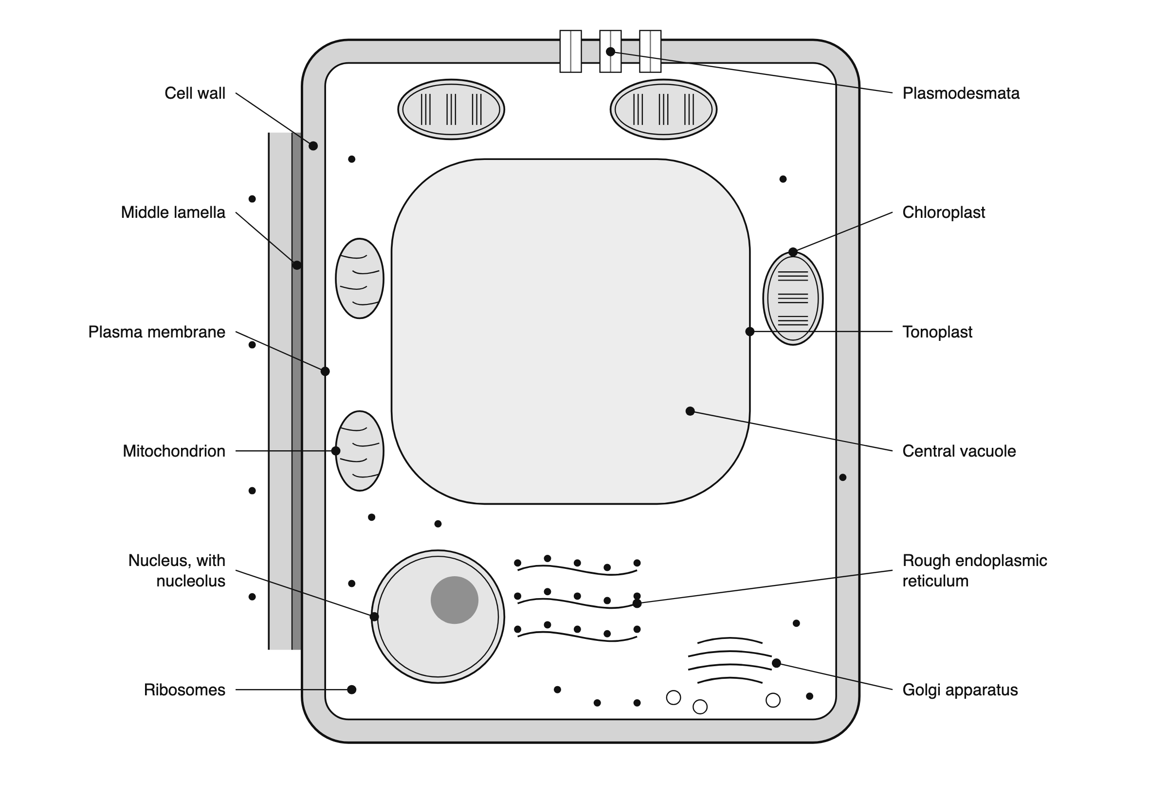
A plant cell diagram is a labelled section through one cell. Working outward from the centre: central vacuole bounded by the tonoplast, nucleus with nucleolus, rough ER, Golgi apparatus, mitochondria, chloroplasts, ribosomes in the cytosol, then the plasma membrane, the cell wall, and the middle lamella joining that wall to the next cell’s, crossed by plasmodesmata. The four an animal cell does not have are the wall, the plastids, the large central vacuole and the plasmodesmata.
What does a plant cell diagram show?
It shows a section. A plant cell is a three-dimensional box, and the diagram cuts it open so the interior is visible — which means the outline you are looking at is a cut edge, not a silhouette.
Three things on the diagram are claims. Which compartments are present. How they nest inside one another: the tonoplast is inside the plasma membrane, which is inside the wall. And which membranes are continuous with which, which is the part almost every diagram draws and almost no reader is told to look for.
Everything else is a drawing decision. The colours are conventional, not observed. The number of chloroplasts drawn is whatever fitted. The small organelles are drawn far larger relative to the cell than they are, because at true relative scale a ribosome in an 84 mm cell would be smaller than the line weight used to draw it.
There is a historical reason the wall dominates the picture. It was the thick cell walls of cork, visible in a primitive microscope, that let Robert Hooke distinguish and name cells for the first time in 1663. What he saw and named was not the living contents; it was the empty boxes their walls left behind.
The parts of a plant cell, labelled
Each label, and what the structure behind it actually is:
| Labelled part | What it is | What it does |
|---|---|---|
| Cell wall | An extracellular matrix of cellulose microfibrils cross-linked by glycans and embedded in a pectin network. Secondary walls add lignin | Bears the tension that lets the cell hold turgor pressure; gives the plant its skeleton; helps form the channels fluid moves through |
| Middle lamella | The pectin-rich layer cementing the walls of two neighbouring cells together | Holds cells in place, which is why plants need no anchoring junctions. Its regulated breakdown is what ripens a tomato and drops a leaf in autumn |
| Plasma membrane | The lipid bilayer against the inner face of the wall | Controls what enters and leaves; carries the cellulose synthase complexes that spin the wall outward from the cell surface |
| Central vacuole | One or several large fluid-filled compartments, typically over 30% of cell volume and up to 90% in some cell types | Storage, digestion, cheap bulk, and control of turgor pressure |
| Tonoplast | The membrane bounding the vacuole | Holds the osmotic gradient that turgor depends on; its transporters are what let a cell hold pressure steady while its surroundings change |
| Nucleus | A double-membraned envelope enclosing the chromosomes, with a nucleolus inside it | Holds the nuclear genome, which encodes about 90% of chloroplast proteins as well as its own |
| Chloroplast | A plastid 5–10 μm long with a double-membrane envelope and a third internal membrane system, the thylakoid membrane, stacked into grana | Photosynthesis; also amino acid and fatty acid synthesis, and the reduction of nitrite to ammonia |
| Mitochondrion | A double-membraned organelle whose inner membrane is folded into cristae | Respiration-driven ATP synthesis. Plant cells have them too — photosynthesis does not replace respiration |
| Rough endoplasmic reticulum | Membrane cisternae studded with ribosomes on the cytosolic face | Synthesis and folding of proteins entering the secretory pathway |
| Golgi apparatus | A stack of flattened cisternae with vesicles budding from its faces | Modifies and sorts proteins and, in plants, supplies most of the non-cellulose wall polysaccharides |
| Ribosomes | Free in the cytosol, bound to the rough ER, and present again inside chloroplasts and mitochondria | Translation. The separate sets inside the organelles are the visible remnant of their bacterial ancestry |
| Plasmodesma | A cytoplasmic channel 20–40 nm across crossing the wall, with a desmotubule of smooth ER running down its centre | The only intercellular junction plants have; passes molecules below about 800 daltons, and selected proteins and RNAs above that |
Two of these repay a second look, because the diagram usually gets their proportions wrong.
The central vacuole is not a storage bag off to one side. Alberts puts it at more than 30% of cell volume, and as much as 90% in some cell types. That number is the reason a plant cell can be large and cheap at the same time: the cytoplasm is a thin shell, and the bulk is water under pressure. It is also doing four jobs simultaneously — storage of nutrients and waste, digestion with hydrolytic enzymes that make it a relative of the animal lysosome, bulk, and control of turgor. Different vacuoles with distinct functions are often present in the same cell.
The chloroplast is not a mitochondrion that happens to be green. Both have a double-membrane envelope, both make ATP chemiosmotically, both descend from endocytosed bacteria. What separates them is the third membrane: the thylakoid membrane, which forms flattened discs stacked into grana and carries the electron-transport chains and the ATP synthase. The chloroplast's inner envelope membrane, unlike the mitochondrion's, is not folded into cristae and plays no part in photosynthesis. A chloroplast drawn as a plain green oval has omitted the only structure that makes it a chloroplast.
What do plant cells have that animal cells do not?
Four structures, and one difference in how the cell divides.
| Structure | Plant cell | Animal cell |
|---|---|---|
| Cell wall | Present. Cellulose, cross-linking glycans, pectin, and lignin in secondary walls — polymers containing no nitrogen | Absent. Animal extracellular matrix is rich in protein and other nitrogen-containing polymers |
| Plastids | Present in all living plant cells. Chloroplasts, chromoplasts, amyloplasts and the rest all develop from proplastids | Absent |
| Large central vacuole | Present, typically over 30% of cell volume | Absent. Animal cells have small vesicles and lysosomes instead |
| Intercellular junctions | Plasmodesmata only | Gap junctions, plus tight, anchoring and other junction classes |
| Cytokinesis | A phragmoplast builds a new wall outward from inside the cell | A contractile ring of actin and myosin pinches the cell in two |
| Mitochondria, nucleus, ER, Golgi, ribosomes | Present | Present |
The junction row is the one worth dwelling on, because it explains the wall. Plant cells are immobilised inside rigid walls, and the walls of adjacent cells are cemented to each other — so plants need no anchoring junctions to hold tissue together. What they still need is communication, and a wall at least 0.1 μm thick is far too thick for a gap junction to span. Plasmodesmata are the answer: with a few specialised exceptions, every living cell in a higher plant is connected to its living neighbours by them.
That has a consequence most diagrams never state. Because the plasma membrane of one cell is continuous with its neighbour's at every plasmodesma, and their cytoplasm is connected through the channel, the cells of a plant can be viewed as forming a syncytium — many nuclei sharing a common cytoplasm.
The wall between two cells is three layers, not one
Draw a plant cell alone on a white page and the wall has nothing to be a wall against. Draw the neighbour, and three things become visible at once.
The middle lamella is a real layer with a job, not a line where two walls happen to touch. It is rich in pectins, cross-linked by calcium, and it is what cements neighbouring walls into a tissue. Its regulated breakdown is the mechanism behind two everyday processes: a tomato ripening, and a leaf detaching in autumn.
The primary wall is thin and extensible, built while the cell is still growing. Once growth stops, many cells deposit a secondary wall in layers inside the primary one, and the commonest additional polymer in it is lignin — which is what makes xylem vessels and fibre cells woody. Both walls are built from the outside of the plasma membrane: cellulose is not made in the ER and secreted like most matrix molecules, it is spun out directly from cellulose synthase complexes embedded in the plasma membrane, and cortical microtubules inside the cell steer the direction they travel.
That last mechanism is why the wall belongs on a diagram at all. The cellulose microfibrils in an elongating cell usually lie perpendicular to the axis of elongation, and their orientation is a large part of what decides the cell's final shape. Because plant cells cannot move, the shape of the whole plant comes down to controlling that orientation, cell by cell.
How do you draw a plant cell diagram?
Build it in the order the structures constrain each other, not in the order they appear in a textbook list.
Start with the wall, and draw it as a band. A single stroke says the wall is a boundary. A band with thickness says it is a compartment — which is what it is, and it is where the plasma membrane goes: against the band's inner face, not floating somewhere inside the cytoplasm.
Put the vacuole in second, and let it take roughly half the area. Everything else has to fit around it, and a vacuole drawn small forces every other organelle into the wrong place. If your cytoplasm looks like a generous open space, the diagram is describing a young meristematic cell rather than the mature one it is captioned as.
Push the rest into the shell the vacuole leaves. The nucleus, being the largest of what remains, decides where the others go. Chloroplasts and mitochondria distribute around the periphery, which is also where they are in a living cell.
Draw chloroplasts with thylakoid stacks. Three or four short stacked lines inside the envelope is enough. Without them a chloroplast and a mitochondrion are the same drawing in two colours, and colour is the one channel that fails in greyscale printing and for colour-blind readers.
Then place the labels, which is the half of the job that gets rushed. The rules are short:
- One leader line per label, straight, ending in a dot on the structure it names. A leader that stops in empty space near the structure is ambiguous.
- No leader crosses another. If two cross, the labels are in the wrong order — swap them rather than routing around.
- Labels outside the cell, ranged in two columns. Text inside the cytoplasm competes with the thing it is describing and cannot be reflowed for a different column width.
- Set lettering at the size the destination requires. For Nature, all text other than panel letters sits between 5 pt minimum and 7 pt maximum, in a sans-serif face, preferably Helvetica or Arial, and the same face across every figure in the paper. Panel letters are 8 pt bold, upright, lower case.
- Draw at the width it will be printed. Nature's standard figure widths are 89 mm single column and 183 mm double column, with a column-and-a-half option at 120–136 mm and a full page depth of 247 mm. A cell diagram drawn on a slide and dropped into a manuscript arrives at a third of the size it was designed for, and 7 pt lettering becomes 2 pt.
- Keep it vector. Nature asks for AI, EPS or PDF for line art and asks specifically that line art and text not be rasterised. A cell diagram is line art in its entirety.
The general rules behind these are the same ones in our guide to making scientific figures, and the Nature Portfolio numbers in full are in the Scientific Reports figure requirements, which covers what happens when a journal declines to publish a specification at all.
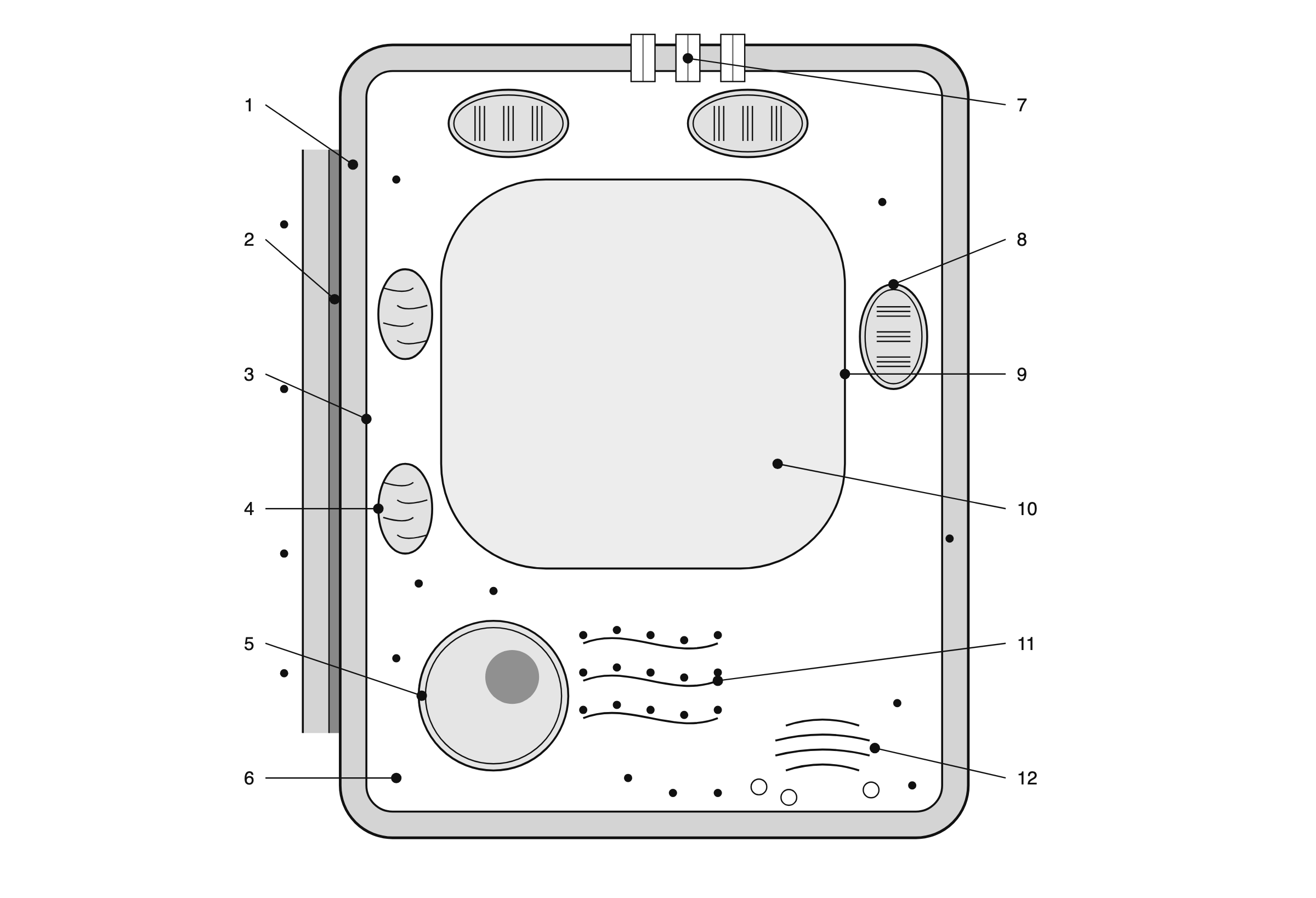
The same diagram without the labels
The most useful version of a diagram you are trying to learn is the one that will not tell you the answer. Same geometry, numbers in place of names.
The key, in the order the numbers run: 1 cell wall, 2 middle lamella, 3 plasma membrane, 4 mitochondrion, 5 nucleus with nucleolus, 6 rough endoplasmic reticulum, 7 plasmodesmata, 8 chloroplast, 9 tonoplast, 10 central vacuole, 11 Golgi apparatus, 12 ribosomes.
Both versions as printable images, at a resolution that holds up on paper:
Five things plant cell diagrams get wrong
The vacuole is drawn too small. This is the commonest error and it propagates, because each new diagram is drawn from the last one. A vacuole occupying a fifth of the cell cannot be the structure that holds a plant upright.
Chloroplasts have no thylakoids. Drawn as flat green ovals they carry no more information than a colour, and the diagram loses the one feature that distinguishes the organelle.
The cell is alone on the page. A wall with nothing on the other side of it invites the reading that the wall is the cell's own skin. It is shared infrastructure: two walls and a middle lamella between two cells, crossed by plasmodesmata. Drawing even a sliver of the neighbour fixes this.
A section is read as an outline. The rounded rectangle is a cut edge through a box. The cytoplasm is a shell surrounding the vacuole on every side, not a ring around a disc. This is the misreading a 3D cutaway is drawn to prevent, at the cost of being much harder to label.
Leader lines cross, or stop short. Both are the same failure — the reader cannot tell which label belongs to which structure — and both are invisible to the person who drew it, because they already know the answer. Check the leaders last, on a printout at final size.
Frequently asked questions
What is a plant cell diagram? A labelled section through a single plant cell, drawn to show which compartments exist and what encloses what. It is a cutaway, not a photograph. The presence of structures, their nesting, and which membranes are continuous are claims; the colours, the counts and the relative sizes of small organelles are drawing decisions.
What are the parts of a plant cell? Cell wall, middle lamella, plasma membrane, cytosol with free ribosomes, nucleus and nucleolus, rough endoplasmic reticulum, Golgi apparatus, mitochondria, chloroplasts and other plastids, and the central vacuole bounded by the tonoplast. Plasmodesmata cross the wall and connect one cell's cytoplasm to the next.
What do plant cells have that animal cells do not? The cell wall, plastids, the large central vacuole and plasmodesmata. Plant cells also divide by building a new wall with a phragmoplast rather than pinching in two with a contractile ring.
What does the central vacuole do? Storage, digestion, bulk, and turgor. Turgor is the load-bearing one: the pressure it holds against the wall drives cell expansion during growth and supplies much of the mechanical rigidity of living plant tissue.
How do you draw a simple plant cell diagram? Wall as a band, vacuole in next at roughly half the area, everything else pushed into the shell that leaves, chloroplasts with visible thylakoid stacks, then straight non-crossing leader lines ending in a dot on each structure.
Is a plant cell diagram 2D or 3D? Nearly always a 2D section through a 3D cell. A 3D version draws the cell as a cutaway solid, which shows the geometry better and is harder to label inside a journal column.
What is the plant cell diagram for class 9? The same section with a reduced label set — wall, membrane, cytoplasm, nucleus, nucleolus, vacuole, chloroplast, mitochondrion, ER, Golgi and ribosomes. The middle lamella and plasmodesmata are usually left off, which is a shame, because they are what make the wall make sense.
Where to go next
- The animal cell diagram, labelled the same way — the cell on the facing page, including the measured volumes that show why almost every diagram draws the nucleus too big, and how two animal cells connect where a plant uses a plasmodesma.
- How to make scientific figures — the general rules behind the labelling section above, applied to every figure type rather than this one.
- Scientific Reports figure requirements — the Nature Portfolio numbers in full, and what to do when a journal publishes no specification at all.
- Cladogram vs phylogenetic tree — another diagram where the layout is routinely mistaken for the claim, and the same discipline of separating what the drawing states from what it merely arranges.
Riferimenti
- Alberts B, et al. Molecular Biology of the Cell, 4th edition — The Plant Cell Wall — Garland Science, via NCBI Bookshelfhttps://www.ncbi.nlm.nih.gov/books/NBK26928/Consultato il 19 ago 2026
- Alberts B, et al. Molecular Biology of the Cell, 4th edition — Cell Junctions — Garland Science, via NCBI Bookshelfhttps://www.ncbi.nlm.nih.gov/books/NBK26857/Consultato il 19 ago 2026
- Alberts B, et al. Molecular Biology of the Cell, 4th edition — Transport from the Trans Golgi Network to Lysosomes — Garland Science, via NCBI Bookshelfhttps://www.ncbi.nlm.nih.gov/books/NBK26844/Consultato il 19 ago 2026
- Alberts B, et al. Molecular Biology of the Cell, 4th edition — Chloroplasts and Photosynthesis — Garland Science, via NCBI Bookshelfhttps://www.ncbi.nlm.nih.gov/books/NBK26819/Consultato il 19 ago 2026
- Alberts B, et al. Molecular Biology of the Cell, 4th edition — An Overview of M Phase — Garland Science, via NCBI Bookshelfhttps://www.ncbi.nlm.nih.gov/books/NBK26931/Consultato il 19 ago 2026
- Cooper GM. The Cell: A Molecular Approach, 2nd edition — Chloroplasts and Other Plastids — Sinauer Associates, via NCBI Bookshelfhttps://www.ncbi.nlm.nih.gov/books/NBK9905/Consultato il 19 ago 2026
- Nature — Final submission: figure preparation — Springer Naturehttps://www.nature.com/nature/for-authors/final-submissionConsultato il 19 ago 2026
I nomi di riviste, congressi e prodotti sono marchi dei rispettivi titolari. Questa pagina è contenuto editoriale indipendente e non è affiliata, autorizzata né approvata da essi. Le specifiche cambiano: verifica ogni requisito sulle fonti ufficiali collegate sopra prima di inviare o stampare.