
Synapse Diagram Maker
Describe the diagram you need and get a labeled chemical synapse — axon terminal, synaptic vesicles, neurotransmitter, voltage-gated Ca²⁺ channels, synaptic cleft, receptors and reuptake transporter. Or view the labeled and blank versions below, free.
Synapse diagram examples
Real renders of the prompts shown: a fully labeled chemical synapse, a five-label school version, a labeled neuromuscular junction and a blank worksheet. Click one to load and edit its prompt.
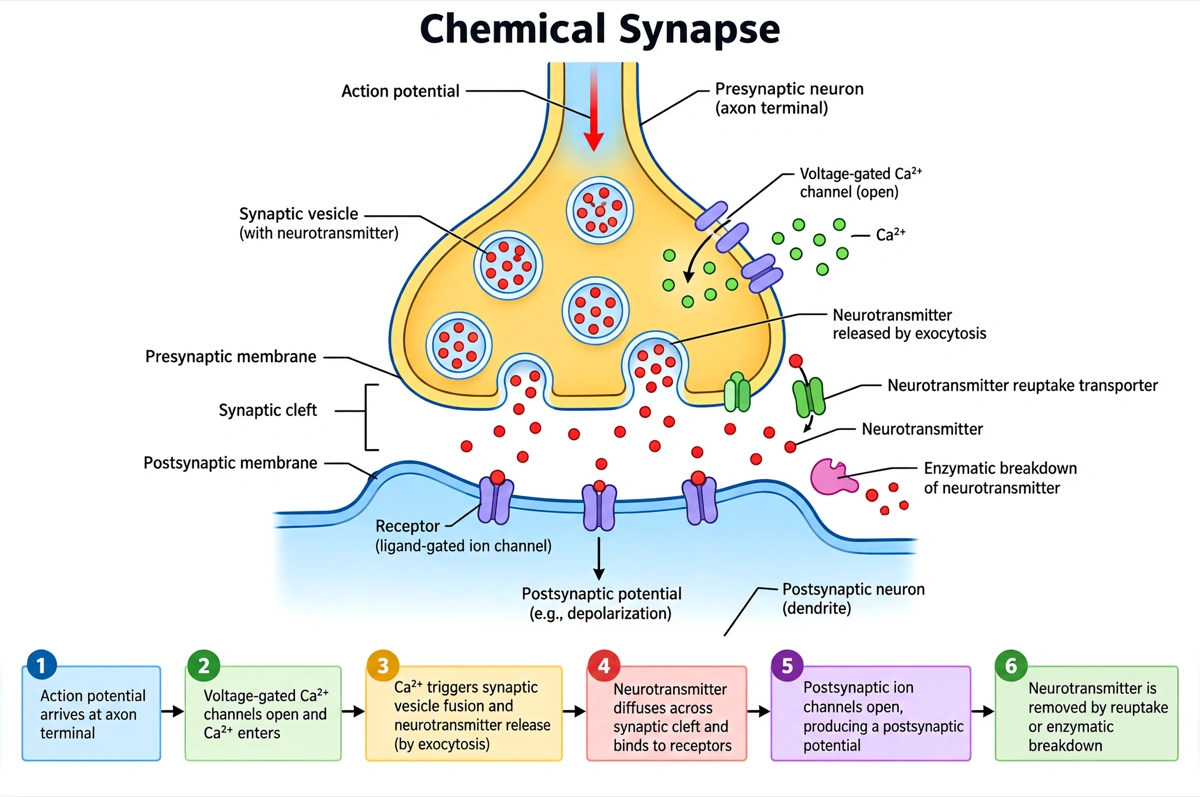
Labeled synapse diagram (answer key)
Every part of a chemical synapse named, from the vesicles in the axon terminal to the receptors on the postsynaptic membrane. Print it as the answer key, or hand it out as a revision sheet.
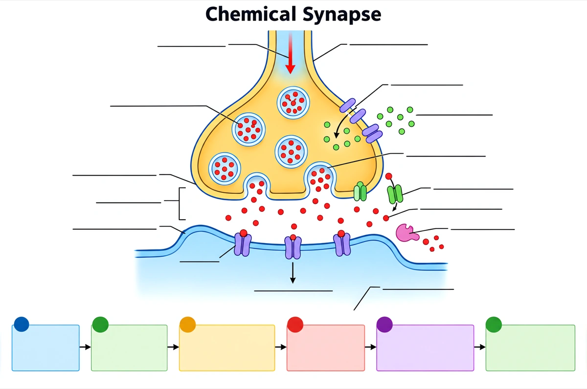
Blank synapse diagram to label
The same drawing with an empty answer line where every label was, ready for a worksheet, a quiz or an exam question.
What is a synapse diagram?
A synapse diagram shows the junction where one neuron passes its signal to the next cell: an action potential opens Ca²⁺ channels in the axon terminal, synaptic vesicles release neurotransmitter into the synaptic cleft, and the neurotransmitter binds to receptors on the postsynaptic membrane. Each part is labeled with its job.
Key facts
- The synaptic cleft is only about 20–40 nm wide. The neurotransmitter crosses it by diffusion, and release plus diffusion add a synaptic delay of about 1 ms between the action potential arriving and the postsynaptic channels opening.
- Release is triggered by Ca²⁺. When the action potential depolarizes the axon terminal, voltage-gated Ca²⁺ channels open, Ca²⁺ flows in, and synaptic vesicles fuse with the presynaptic membrane and empty by exocytosis.
- A chemical synapse works in one direction only. Vesicles sit in the presynaptic terminal and receptors on the postsynaptic membrane, so the signal cannot run backwards — unlike some electrical synapses, which pass current both ways through gap junctions.
- The response depends on the receptor. Ionotropic receptors are ligand-gated ion channels that open within milliseconds; metabotropic receptors act through G proteins and second messengers, more slowly and for longer.
- The signal is ended in three ways: enzymes, reuptake and diffusion. Acetylcholinesterase splits acetylcholine in the cleft; serotonin, dopamine and norepinephrine are pumped back into the terminal by reuptake transporters, the target of SSRIs.
How does the synapse diagram maker work?
Describe the diagram
Say what it is for and how deep it should go — a full labeled chemical synapse, a simple five-label overview, a neuromuscular junction, a chemical and an electrical synapse side by side. A sentence is enough.
Pick a style
The default is a clean flat textbook plate — white background, labels on straight leader lines. Or apply watercolor, ink line art, 3D and other finishes.
Refine and export
The result opens in your workspace, where you can redraw regions, edit labels on the canvas, upscale and export in high resolution.
Parts of a synapse and what each one does
The parts a labeled chemical synapse diagram is normally expected to show, what each one does, and where it sits. This is the default label set unless you ask for a shorter one.
| Part | What it does | Where it sits |
|---|---|---|
| Axon terminal (presynaptic terminal) | Turns the arriving action potential into a chemical signal | End of the presynaptic neuron's axon |
| Synaptic vesicles | Store neurotransmitter and release it by exocytosis | Inside the axon terminal, clustered at the active zone |
| Neurotransmitter | The chemical messenger — acetylcholine, glutamate, GABA, dopamine and others — that carries the signal across the cleft | Stored in vesicles, released into the cleft |
| Voltage-gated Ca²⁺ channels | Open when the terminal depolarizes and let Ca²⁺ in, which triggers vesicle fusion | Presynaptic membrane, at the active zone |
| Mitochondria | Supply the ATP for refilling vesicles and pumping ions | Inside the axon terminal |
| Synaptic cleft | The gap, about 20–40 nm wide, that the neurotransmitter diffuses across | Between the presynaptic and postsynaptic membranes |
| Receptors (ligand-gated ion channels) | Bind the neurotransmitter and open, letting ions through to change the membrane potential | Postsynaptic membrane |
| Postsynaptic membrane | Carries the receptors; its change in potential is the EPSP or IPSP | Dendrite, cell body or muscle fiber of the receiving cell |
| Reuptake transporter | Carries neurotransmitter back into the terminal, ending the signal and recycling the transmitter | Presynaptic membrane and nearby glial cells |
| Enzymes in the cleft (such as acetylcholinesterase) | Break the neurotransmitter down | Synaptic cleft |
Courses differ in how much of the synapse they expect. Introductory worksheets usually stop at the axon terminal, vesicles, neurotransmitter, synaptic cleft and receptors; A-level, AP and university courses add the Ca²⁺ channels, reuptake or enzyme breakdown, mitochondria and the split between ionotropic and metabotropic receptors. Name the labels you need and only those are drawn.
Chemical vs electrical synapse: what is the difference?
A chemical synapse passes the signal as a neurotransmitter released across a cleft of about 20–40 nm; an electrical synapse passes ions straight from cell to cell through gap junctions. Chemical synapses are by far the more common; electrical synapses are faster and keep groups of neurons firing in step.
| Chemical synapse | Electrical synapse | |
|---|---|---|
| Gap between the cells | Synaptic cleft, about 20–40 nm | Gap junction, about 3.5 nm, the membranes joined by channel proteins |
| How the signal crosses | Neurotransmitter released from vesicles binds receptors | Ions flow directly through the gap-junction channels |
| Speed | Synaptic delay of about 1 ms | Virtually instantaneous |
| Direction | One-way, presynaptic to postsynaptic | Often two-way |
| Effect on the signal | Can excite or inhibit, amplify, and be changed by drugs | Passes the change in voltage on with little alteration |
| Plasticity | Strengthened or weakened by use — long-term potentiation and depression, the basis of learning | Little; mainly synchronizes the activity of neighboring cells |
Draw the chemical synapse with a visible cleft, vesicles on the presynaptic side only and receptors on the postsynaptic side only, and the electrical synapse with the two membranes almost touching, joined by gap-junction channels. Receptors on both sides of a chemical synapse, or vesicles in an electrical one, are the errors to check for first.
Where are synapse diagrams used?
Synapse diagrams are used mostly in biology and physiology teaching — labeling worksheets, exam revision and lecture slides — and in medicine and pharmacology, where many drugs that act on the nervous system are explained at the synapse. What changes is the depth: five labels for a first course, every channel and transporter for a medical one.
School biology
Labeling worksheets and revision sheets for GCSE, A-level, AP Biology and IB, where the synapse comes with nervous coordination and the effect of drugs. Usually the labeled synapse, plus the same drawing blank for students to fill in.
Anatomy, physiology and nursing
A&P and nursing slides on how a nerve signal passes from cell to cell and onto muscle, with the neuromuscular junction for the chapter on skeletal muscle contraction.
Medicine and pharmacology
Slides that put each drug on its step: SSRIs on serotonin reuptake, acetylcholinesterase inhibitors in the cleft, botulinum toxin on vesicle release, neuromuscular blockers on the acetylcholine receptors.
Research and textbook figures
Schematic panels for neuroscience papers, grant applications and review articles — a synapse with the protein of interest highlighted, drawn at the depth the figure needs.
Common questions about synapse diagrams
What are the parts of a synapse?
A chemical synapse has three parts: the presynaptic axon terminal, the synaptic cleft and the postsynaptic membrane. The terminal holds synaptic vesicles of neurotransmitter, mitochondria and voltage-gated Ca²⁺ channels; the postsynaptic membrane carries the receptors. A labeled diagram usually adds the neurotransmitter molecules and a reuptake transporter or an enzyme in the cleft.
What are the steps of synaptic transmission?
An action potential reaches the axon terminal; voltage-gated Ca²⁺ channels open and Ca²⁺ flows in; vesicles release neurotransmitter by exocytosis; it diffuses across the cleft; it binds receptors that open ion channels in the postsynaptic membrane; and it is removed by enzymes, reuptake or diffusion. The chemical step from release to response takes about 1 ms.
What is the difference between a chemical and an electrical synapse?
A chemical synapse passes the signal with a neurotransmitter across a cleft of about 20–40 nm; an electrical synapse lets ions flow straight through gap junctions between two cells that almost touch. Chemical synapses are one-way, about 1 ms slower and adjustable; electrical synapses are virtually instantaneous and often two-way.
What is the neuromuscular junction?
The neuromuscular junction is the chemical synapse between a motor neuron and a skeletal muscle fiber. The axon terminal releases acetylcholine, which binds nicotinic acetylcholine receptors on the motor end plate and starts an action potential in the muscle fiber; acetylcholinesterase in the cleft then breaks the acetylcholine down.
Can I get a blank synapse diagram to label?
Yes — view the blank version on this page free, or ask for one and every label is replaced by an empty answer line. The labeled version is the matching answer key, so the two print as a pair.
What does the synaptic cleft do?
It keeps the two cells apart, so the signal has to cross as a chemical — which is what lets a synapse amplify, inhibit or block it. The cleft is about 20–40 nm wide; the neurotransmitter diffuses across it, and enzymes and transporters at its edges clear it away.
Why is calcium needed for neurotransmitter release?
Ca²⁺ entering the axon terminal is the trigger that makes synaptic vesicles fuse with the presynaptic membrane. Without it the action potential still arrives but no neurotransmitter is released, which is why blocking the voltage-gated Ca²⁺ channels stops transmission.
How is a neurotransmitter removed from the synapse?
By enzyme breakdown, by reuptake into the presynaptic terminal or nearby glial cells, or by diffusing out of the cleft. Acetylcholinesterase splits acetylcholine; SSRI antidepressants block serotonin reuptake, so serotonin stays in the cleft longer.
What is the difference between an excitatory and an inhibitory synapse?
An excitatory synapse depolarizes the postsynaptic membrane (an EPSP), moving it toward threshold; an inhibitory synapse hyperpolarizes it (an IPSP), moving it away. Glutamate is the main excitatory neurotransmitter in the brain and GABA the main inhibitory one; whether the neuron fires depends on the sum of all its inputs.
How is this different from a generic AI image tool?
The subject brief is built in. Every render is instructed to follow the standard textbook drawing — the axon terminal, cleft and postsynaptic membrane in their true positions and proportions, vesicles only on the presynaptic side — with each label spelled exactly and set on a straight leader line. Check the labels against your course before printing; the canvas lets you correct any in place.
Can I use the diagrams for school, teaching or publication?
Yes — diagrams you generate are yours to use in worksheets, presentations, handouts, theses and papers. Export high-resolution raster up to 4K depending on the model.
Can I change the labels after generating?
Yes. The result opens in a canvas workspace that recognizes the diagram labels — you can edit or translate the text, move leader lines, redraw a region or recolor without regenerating the whole figure.
Keep exploring
Our other diagram tools, and the guides behind them.

Draw your synapse diagram
From a one-line description to a labeled, printable diagram in about a minute.
Start drawing