
Notch Signaling Pathway Diagram Maker
Describe the figure you need and get a labeled Notch signaling pathway between two cells — the ligand, the Notch receptor, the S2 and S3 cuts, NICD entering the nucleus and CSL (RBPJ) switching on HES and HEY genes.
Notch signaling pathway examples
Real renders of four Notch signaling prompts: the fully labeled pathway, a five-label overview, lateral inhibition in the inner ear and a blank worksheet. Click one to load its prompt and edit it.
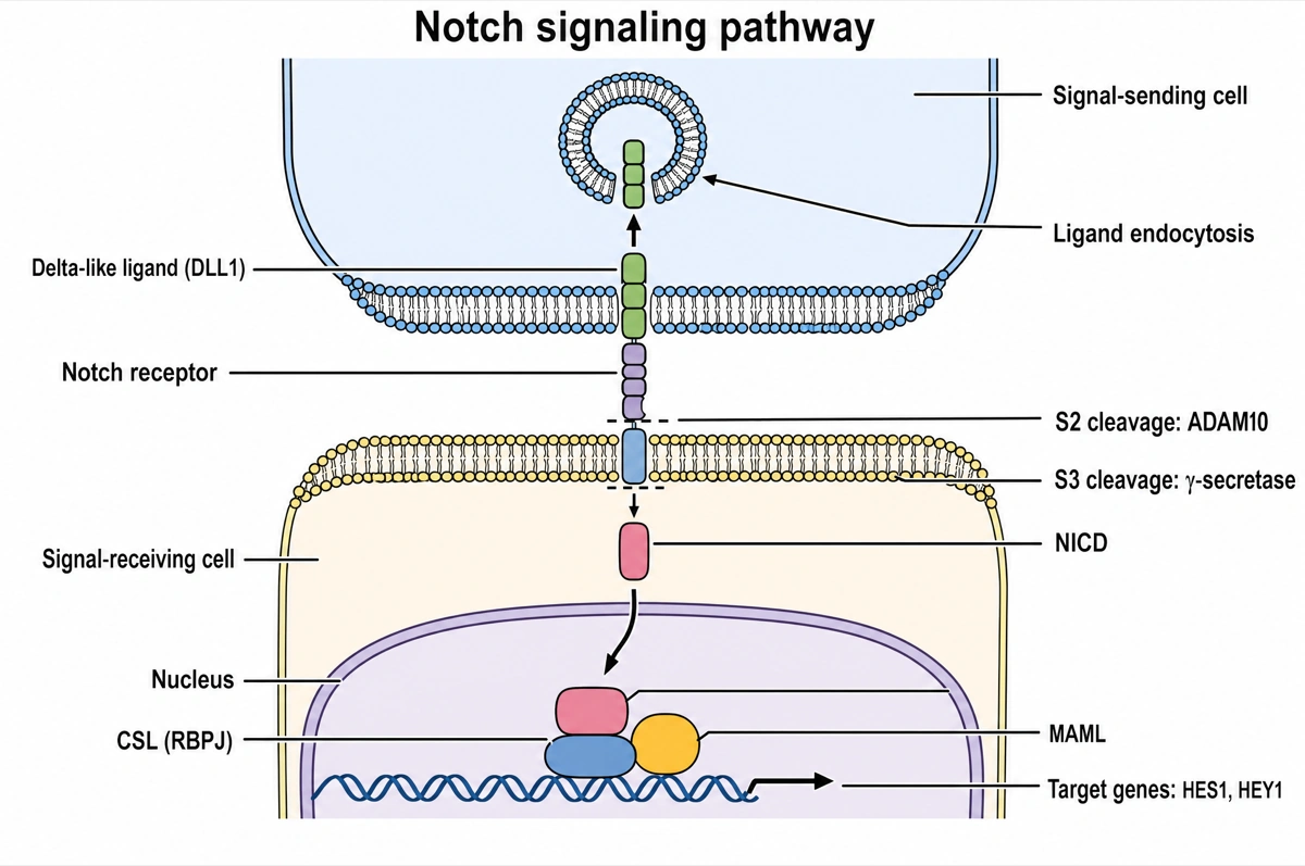
Two cells in contact, a receptor cut twice
Read down from the sending cell to the nucleus below.
- 1
DLL1 → Notch receptor
The ligand on the sending cell binds Notch on the receiving cell.
- 2
Ligand endocytosis
The sending cell pulls the bound ligand in, tugging on Notch.
- 3
S2 and S3 cleavage
ADAM10 cuts outside the membrane; γ-secretase then cuts inside.
- 4
NICD, CSL (RBPJ), MAML
In the nucleus, NICD joins CSL and MAML to turn on HES1 and HEY1.
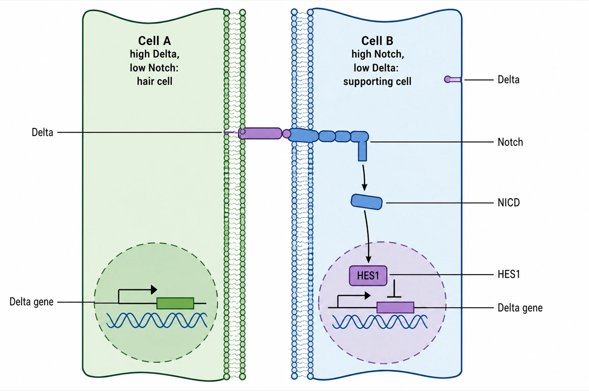
One cell keeps its Delta, the other loses it
Read left to right, from Cell A across into Cell B.
- 1
Delta on A → Notch on B
Cell A, high in Delta, activates Notch on its neighbour, Cell B.
- 2
NICD → HES1
NICD released in Cell B switches on HES1.
- 3
HES1 on B's Delta gene
HES1 turns down B's own Delta, so the difference between them grows.
What is the Notch signaling pathway?
The Notch signaling pathway is a contact-dependent pathway: a Delta-like or Jagged ligand on one cell binds the Notch receptor on its neighbour, ADAM10 and γ-secretase cut the receptor, and the freed Notch intracellular domain (NICD) enters the nucleus to switch on HES and HEY genes with CSL (RBPJ) and MAML. The receptor fragment itself carries the signal, with no kinase cascade or second messenger in between.
Key facts
- Mammals have four Notch receptors and five canonical ligands. NOTCH1–4 are activated by the Delta-like ligands DLL1 and DLL4 and the Jagged ligands JAG1 and JAG2; DLL3 works mainly by cis-inhibition rather than by activating Notch.
- Notch is cut three times. Furin cleaves the new receptor at S1 in the Golgi, making the mature heterodimer; after ligand binding, ADAM10 cuts at S2 just outside the membrane, and γ-secretase, whose catalytic subunit is presenilin, cuts at S3 inside it to release NICD.
- The ligand has to pull. Binding alone is not enough: the ubiquitin ligases Mindbomb or Neuralized tag the bound ligand, the sending cell endocytoses it, and that pull unfolds the receptor's negative regulatory region so ADAM10 can reach the S2 site.
- CSL (RBPJ) is a switch. Without NICD it sits on target genes with co-repressors and histone deacetylases; NICD displaces them and recruits MAML and histone acetyltransferases such as p300 to turn HES1, HES5, HEY genes and MYC on.
- Each signal is short-lived. CDK8 phosphorylates NICD's PEST domain and the ubiquitin ligase FBXW7 marks it for destruction, resetting the pathway — PEST and FBXW7 mutations that stop this are common in T-cell acute lymphoblastic leukemia.
How does the Notch signaling pathway diagram maker work?
Describe the diagram
Say what it is for and how deep it should go — the full two-cell pathway, a simple overview, lateral inhibition between neighbours. A sentence is enough.
Pick a style
The default is a clean flat textbook plate — white background, straight arrows, labels on leader lines. Or apply watercolor, ink line art, 3D and other finishes.
Refine and export
The result opens in your workspace, where you can redraw regions, edit labels on the canvas, upscale and export in high resolution.
Components of the Notch signaling pathway and what each one does
The molecules a labeled Notch pathway diagram is normally expected to show, in the order the signal passes through them. This is the default label set unless you ask for a shorter one.
| Component | What it does | Where to draw it |
|---|---|---|
| DSL ligand (DLL1, DLL4, JAG1, JAG2) | Transmembrane ligand that binds Notch on the neighbouring cell | In the sending cell's membrane, facing the gap |
| Mindbomb or Neuralized | Ubiquitin ligases that tag the bound ligand for endocytosis, which pulls on the receptor | Inside the sending cell, at the ligand's tail |
| Notch receptor (NOTCH1–4) | Single-pass receptor, held as a two-part heterodimer after S1 cleavage by furin | Spanning the receiving cell's membrane |
| Fringe | Golgi glycosyltransferase that extends O-fucose on Notch's EGF repeats, favouring Delta over Jagged | In the receiving cell's Golgi (optional) |
| ADAM10 (S2 cleavage) | Metalloprotease that sheds the extracellular domain once the pull exposes the S2 site, leaving NEXT | Just outside the receiving cell's membrane |
| γ-Secretase (S3 cleavage) | Intramembrane protease — presenilin with nicastrin, PEN-2 and APH-1 — that cuts NEXT to release NICD | Within the membrane bilayer |
| NICD | Notch intracellular domain; carries the signal itself into the nucleus | Arrow from the membrane through the cytoplasm to the nucleus |
| CSL (RBPJ) with co-repressors | DNA-binding protein that keeps target genes off while NICD is absent | On the DNA in the nucleus |
| MAML (Mastermind-like) | Co-activator recruited by NICD and CSL; brings in p300 | Stacked on NICD and CSL on the DNA |
| HES and HEY genes | Main targets; they encode repressors that carry out Notch's effects | Downstream of the complex, at the bottom |
Courses differ in how many of these they expect. An introductory figure stops at ligand, receptor, NICD and target genes; a cell biology course adds ADAM10, γ-secretase, CSL (RBPJ) and MAML; advanced courses add furin, Fringe and Mindbomb. Sources also differ on the S2 enzyme: ADAM17 cuts Notch in the test tube, but ligand-induced cleavage in living cells depends on ADAM10.
Notch vs Wnt signaling: what is the difference?
Notch signaling needs direct contact — a membrane-bound ligand on one cell triggers cuts in the receptor on the next, and the receptor fragment itself goes to the nucleus — whereas canonical Wnt signaling uses a secreted ligand that saves β-catenin from its destruction complex. Both steer cell fate in development and stem cells, and both are rewired in cancer.
| Notch signaling | Wnt (canonical) signaling | |
|---|---|---|
| Ligand | Membrane-bound DSL ligands: DLL1, DLL4, JAG1, JAG2 | Secreted, lipid-modified WNT glycoproteins — 19 in humans |
| Range | Contact-dependent (juxtacrine): only touching cells | Short and long range, as a gradient around the source |
| Receptor | NOTCH1–4, single-pass transmembrane | One of 10 Frizzled receptors with the LRP5/6 co-receptor |
| How the signal passes | Proteolysis: S2 by ADAM10, S3 by γ-secretase | The destruction complex (APC, Axin, GSK3, CK1) is switched off |
| Messenger to the nucleus | NICD, a fragment of the receptor | β-catenin, a cytoplasmic protein spared from degradation |
| Nuclear partner | CSL (RBPJ) with MAML | TCF/LEF |
| Off state | CSL (RBPJ) represses targets with co-repressors | β-catenin is phosphorylated and destroyed by the proteasome |
| Signature cancer | NOTCH1 mutations in more than 50% of T-ALL | A Wnt-pathway mutation in about 90% of sporadic colorectal cancers |
Draw Notch as two cells touching, with the ligand fixed in one membrane; draw Wnt with the ligand free in the space between cells. The two pathways cross-talk — NICD can act on Wnt signaling — but keep their nuclear partners apart: CSL for Notch, TCF/LEF for Wnt.
Where are Notch signaling pathway diagrams used?
Notch pathway diagrams are used in cell and developmental biology teaching, in exam revision and as schematic panels in stem-cell, vascular and cancer research. The depth changes with the audience: four labels for a first course, every cleavage and co-factor for a graduate one.
Cell biology courses
Lecture slides on cell–cell signaling that set Notch beside Wnt, Hedgehog and TGF-β: a membrane-bound ligand, activation by proteolysis and a receptor fragment that works as a transcription co-activator.
Developmental biology
Figures of lateral inhibition — hair cells and supporting cells in the inner ear, tip and stalk cells in growing blood vessels — and of somite segmentation, where pulses of Notch activity mark each new somite.
Cancer and hematology
Panels on NOTCH1 activating mutations in T-ALL, FBXW7 loss, and drugs that act on the pathway, from γ-secretase inhibitors to DLL3-targeted antibodies in small-cell lung cancer.
Research figures and grant proposals
Schematics that mark where a mutation, a knockout or an inhibitor acts — ADAM10, γ-secretase, CSL (RBPJ) or MAML — directly on the pathway.
Common questions about the Notch signaling pathway
What should a labeled Notch signaling pathway diagram include?
The standard set is the signal-sending cell with a DSL ligand (Delta-like or Jagged), the signal-receiving cell with the Notch receptor, S2 cleavage by ADAM10, S3 cleavage by γ-secretase, the released NICD, and in the nucleus CSL (RBPJ), MAML and the HES/HEY target genes. Many figures add S1 cleavage by furin in the Golgi and the co-repressors CSL holds before NICD arrives.
What are the steps of Notch signaling?
Five steps: a ligand on one cell binds Notch on the next; the sending cell endocytoses the ligand and pulls; ADAM10 cuts Notch at S2; γ-secretase cuts it at S3 inside the membrane; and NICD enters the nucleus, where it joins CSL (RBPJ) and MAML to switch on target genes. Before any of this, furin has already cut the new receptor once, at S1 in the Golgi.
What does γ-secretase do in Notch signaling?
γ-Secretase makes the last cut, S3, inside the membrane, releasing NICD from the stub that ADAM10 leaves behind. It is a four-part complex — presenilin 1 or 2 as the catalytic subunit, with nicastrin, PEN-2 and APH-1 — and it also cleaves amyloid precursor protein, the substrate linked to Alzheimer's disease.
What is lateral inhibition in Notch signaling?
Lateral inhibition is how Notch splits a group of equivalent cells into two fates: a cell that sends slightly more Delta activates Notch in its neighbours, Notch signaling lowers their own Delta, and the difference grows until the first cell takes one fate and the cells around it another. In the inner ear this leaves each hair cell surrounded by supporting cells.
What is cis-inhibition of Notch?
Cis-inhibition is Notch being blocked by ligand in its own cell: a Delta or Jagged ligand in the same membrane binds the receptor and stops it responding to ligand on neighbours. DLL3 seems to act only this way, and together with activation in trans it pushes a cell into a clear sending or receiving state.
What does Fringe do to Notch?
Fringe enzymes — Lunatic, Manic and Radical Fringe — extend the O-fucose sugars on Notch's EGF repeats in the Golgi, which makes NOTCH1 respond more to Delta-like ligands than to Jagged. The modification tunes the signal; it is not required for the receptor to mature.
How is Notch signaling involved in cancer?
Activating NOTCH1 mutations occur in more than 50% of T-cell acute lymphoblastic leukemias (T-ALL), mostly in the heterodimerization domain, which lets the receptor fire without ligand, and in the PEST domain, which keeps NICD from being degraded. In other tissues, such as squamous cell carcinomas, Notch acts as a tumor suppressor instead.
What are γ-secretase inhibitors used for?
γ-Secretase inhibitors (GSIs) block the S3 cut and so shut Notch signaling down; the FDA approved one, nirogacestat, for progressing desmoid tumors in November 2023. An early GSI trial in T-ALL was halted by severe diarrhea — the gut lining depends on Notch, and the enzyme has many substrates besides Notch.
How is this different from a generic AI image tool?
The subject brief is built in. Every render is instructed to lay the pathway out top to bottom — sending cell, receiving cell, the cuts, the nucleus — with plain arrows for activation, flat-headed bars for inhibition and every label spelled as in a textbook. Check the labels against your course before printing; the canvas lets you correct any in place.
Can I use the diagrams for teaching or publication?
Yes — diagrams you generate are yours to use in worksheets, presentations, handouts, theses and papers. Export high-resolution raster up to 4K depending on the model.
Can I change the labels after generating?
Yes. The result opens in a canvas workspace that recognizes the diagram labels — you can edit or translate the text, move leader lines, redraw a region or recolor without regenerating the whole figure.
Sources
- Signaling by NOTCH1 — pathway R-HSA-1980143 — Reactome
- Notch signaling pathway — KEGG PATHWAY hsa04330 — KEGG
- Notch signaling pathway: architecture, disease, and therapeutics (Zhou et al., 2022) — Signal Transduction and Targeted Therapy
- FDA approves nirogacestat for desmoid tumors (2023) — U.S. Food and Drug Administration
Keep exploring
Our other diagram tools, and the guides behind them.

Draw your Notch signaling pathway
From a one-line description to a labeled, printable pathway diagram in about a minute.
Start drawing