
Coagulation Cascade Diagram
Describe the clotting cascade you need and get a clean pathway diagram — the intrinsic and extrinsic pathways joining at factor X, the common pathway down to a cross-linked fibrin clot, every activated factor marked with its “a”.
Coagulation cascade diagram examples
Real renders of the prompts shown: the classical coagulation cascade with all three pathways, a six-box lecture overview, the PT vs aPTT arms and a blank version. Click one to load and edit its prompt.
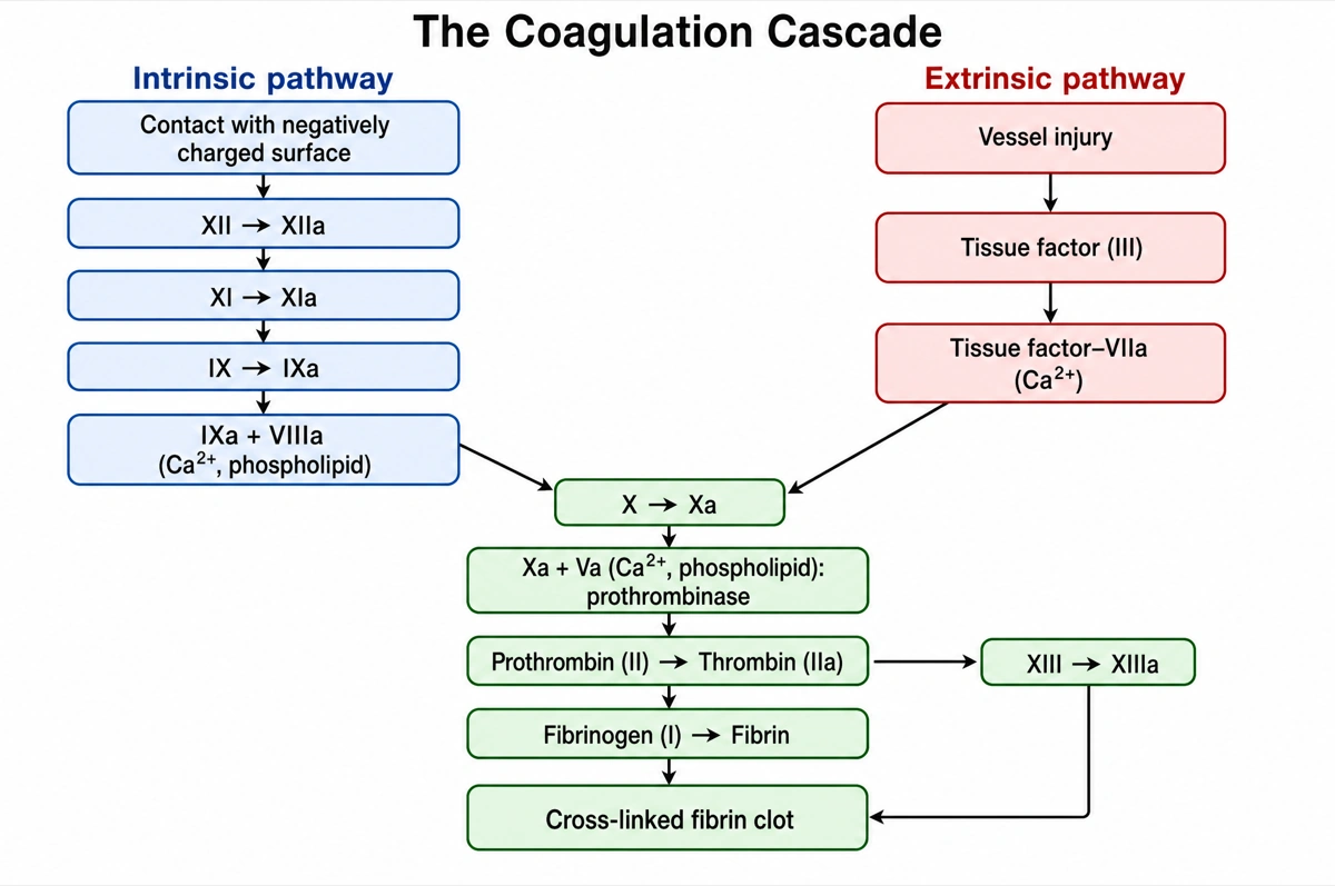
Two arms that meet at factor X
Read both arms to factor X, then the common stem.
- 1
Intrinsic: XII → IXa
Surface contact activates XII, then XI and IX; IXa pairs with VIIIa.
- 2
Extrinsic: tissue factor
Vessel injury exposes tissue factor (III), which binds factor VIIa.
- 3
X → Xa → prothrombinase
Both arms activate factor X; Xa with Va forms prothrombinase.
- 4
Thrombin → fibrin clot
Thrombin cuts fibrinogen into fibrin; XIIIa cross-links the clot.
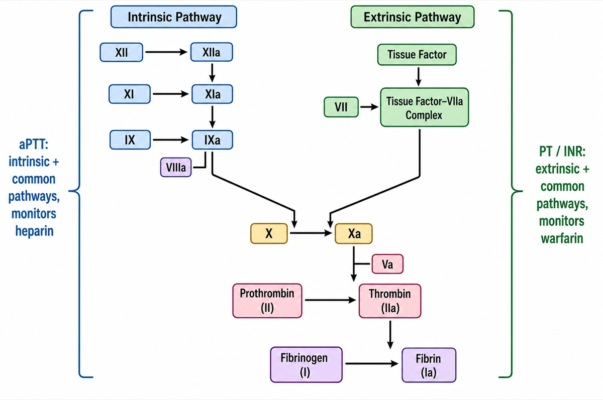
What the PT and the aPTT each measure
Each lab test is bracketed around the arm it covers.
- 1
aPTT: intrinsic + common
XII, XI, IX and VIIIa down to X; the test that monitors heparin.
- 2
PT/INR: extrinsic + common
Tissue factor with VIIa down to X; the test that monitors warfarin.
- 3
Common stem from factor X
Xa and Va, thrombin, fibrin: a defect here prolongs both tests.
What is the coagulation cascade?
The coagulation cascade is the chain of enzyme reactions in which clotting factors — plasma proteins numbered I to XIII — activate one another in turn, from factor XII in the intrinsic pathway or tissue factor with factor VII in the extrinsic pathway, through factor X and thrombin, until soluble fibrinogen becomes a cross-linked fibrin clot. An activated factor carries the suffix “a”: factor X becomes Xa.
Key facts
- Two starting arms meet at factor X. In the extrinsic (tissue factor) pathway, injury exposes tissue factor, which binds factor VIIa; in the intrinsic (contact activation) pathway, XIIa activates XI and XIa activates IX, and IXa with its cofactor VIIIa activates factor X. From Xa onward the steps are shared: the common pathway.
- Thrombin is the enzyme that makes the clot. Factor Xa with its cofactor Va on a phospholipid surface forms prothrombinase, which converts prothrombin (II) into thrombin (IIa); thrombin cuts fibrinogen (I) into fibrin and activates factor XIII, and XIIIa cross-links the fibrin into a stable clot.
- Calcium is factor IV. Ca²⁺ is needed in most thrombin-generating reactions, which is why citrate and EDTA keep a blood sample liquid; the vitamin K–dependent factors II, VII, IX and X bind phospholipid surfaces through calcium bridges.
- There is no factor VI. The factors were numbered I to XIII in order of discovery; factor VI turned out to be the same as factor V, and the number was left empty rather than renumbering the rest.
- In the body the cascade runs as the cell-based model. Initiation on tissue factor–bearing cells makes a little thrombin; amplification uses it to activate platelets and factors V, VIII and XI; propagation on the platelet surface produces a burst of thrombin. The intrinsic/extrinsic split still explains the PT and aPTT tests.
How does the coagulation cascade diagram maker work?
Describe the cascade
Say what you need — the full classical cascade, a six-box overview, the PT and aPTT arms, the sites where warfarin and heparin act, or a blank worksheet. Name the factors you want and only those are drawn.
Pick a style
The default is a clean flat textbook plate: one box per factor, straight arrows from each factor to the one it activates, the three pathways headed and color-coded. Or apply watercolor, ink line art and other finishes.
Refine and export
The result opens in your workspace, where you can redraw regions, edit labels on the canvas, upscale and export in high resolution.
Clotting factors in the coagulation cascade
The factors a labeled coagulation cascade diagram is normally expected to show, their names, what each one does and the pathway it belongs to. This is the default content unless you ask for something else.
| Factor | Name | What it does | Pathway |
|---|---|---|---|
| XII | Hageman factor | Activated to XIIa by contact with a negatively charged surface; activates factor XI | Intrinsic |
| XI | Plasma thromboplastin antecedent | Activated to XIa; activates factor IX. Deficiency causes hemophilia C | Intrinsic |
| IX | Christmas factor | Activated to IXa, the enzyme of the IXa–VIIIa complex that activates factor X. Deficiency causes hemophilia B | Intrinsic |
| VIII | Antihemophilic factor | Activated to VIIIa, the cofactor of IXa. Deficiency causes hemophilia A | Intrinsic |
| III | Tissue factor | Exposed on cells around the vessel wall after injury; binds factor VIIa | Extrinsic |
| VII | Proconvertin | Forms the tissue factor–VIIa complex, which activates factor X | Extrinsic |
| X | Stuart–Prower factor | Activated to Xa, the enzyme of prothrombinase | Common |
| V | Proaccelerin | Activated to Va, the cofactor of Xa in prothrombinase | Common |
| II | Prothrombin | Converted by prothrombinase into thrombin (IIa) | Common |
| I | Fibrinogen | Cut by thrombin into fibrin strands | Common |
| XIII | Fibrin-stabilizing factor | Activated by thrombin to XIIIa, which cross-links fibrin | Common |
| IV | Calcium ions (Ca²⁺) | Needed in most steps; bridges factors to the phospholipid surface | All three |
Factors II, VII, IX and X are vitamin K–dependent, which is why warfarin, a vitamin K antagonist, lowers all four. Name the factors you need and only those are drawn.
Intrinsic vs extrinsic pathway: what is the difference?
The extrinsic pathway starts outside the blood, when injury exposes tissue factor, which binds factor VIIa; the intrinsic pathway starts inside it, when factor XII meets a negatively charged surface and activates XI, then IX, with VIIIa as cofactor. Both end by activating factor X, where the common pathway begins.
| Intrinsic pathway | Extrinsic pathway | |
|---|---|---|
| Other name | Contact activation pathway | Tissue factor pathway |
| Trigger | Factor XII contacting a negatively charged surface, such as glass in a test tube | Tissue factor exposed by damage to the vessel wall |
| Factors | XII, XI, IX and VIII | Tissue factor (III) and VII |
| Complex that activates X | IXa with VIIIa on a phospholipid surface, with Ca²⁺ | Tissue factor–VIIa, with Ca²⁺ |
| Speed | A few minutes | Seconds |
| Lab test | aPTT (activated partial thromboplastin time) | PT (prothrombin time), reported as the INR |
| Inherited deficiency | Hemophilia A (VIII), B (IX) and C (XI) | Factor VII deficiency, which is rare |
| Drug usually monitored | Unfractionated heparin | Warfarin |
Factor XII is not needed for clotting in the body: people born without it have no bleeding problem, although it starts clotting in a glass test tube. That is one reason the cell-based model has replaced the two-arm scheme for describing clotting in vivo.
Where are coagulation cascade diagrams used?
Coagulation cascade diagrams are used mostly in medical, nursing and pharmacy teaching — physiology, hematology and pharmacology — and as figures in hemostasis and thrombosis papers. The version changes with the reader: six boxes for a first lecture, every factor and cofactor for an exam, the cell-based model for research.
Medical and nursing school
Physiology and hematology lectures and exam revision, where the classical cascade with its factor numbers and activated forms is expected by heart, often as a blank diagram to fill in.
Pharmacology
Showing where anticoagulants act: warfarin lowering the vitamin K–dependent factors II, VII, IX and X, heparin boosting antithrombin against thrombin and Xa.
Laboratory medicine
Explaining a prolonged PT or aPTT: which arm of the cascade each test covers and which factor deficiencies or drugs lengthen it.
Research figures and posters
Hemostasis and thrombosis papers, reviews and conference posters, from a simple overview to the initiation, amplification and propagation phases of the cell-based model.
Common questions about the coagulation cascade
How do I draw a coagulation cascade diagram?
Draw the intrinsic pathway (XII → XI → IX, with VIIIa) on one side and the extrinsic pathway (tissue factor with VIIa) on the other, join both arrows at factor X, then run the common pathway down the middle: Xa with Va turns prothrombin into thrombin, thrombin turns fibrinogen into fibrin, and XIIIa cross-links it. With this maker you describe the version you need and the cascade is drawn top to bottom in that order.
Is the clotting cascade the same as the coagulation cascade?
Yes. Clotting cascade, coagulation cascade and blood coagulation pathway all name the same series of factor activations. Ask for a clotting cascade diagram and you get the same figure.
What are the three pathways of the coagulation cascade?
The intrinsic pathway (factors XII, XI, IX and VIII), the extrinsic pathway (tissue factor and factor VII) and the common pathway (factors X, V, II, I and XIII). The first two each activate factor X; everything after that is shared.
Which factors are in the common pathway?
Factor X, its cofactor V, prothrombin (II), fibrinogen (I) and factor XIII, with calcium throughout. Xa and Va form prothrombinase, which makes thrombin; thrombin makes fibrin and activates XIII, which cross-links it.
What is the difference between PT and aPTT?
The PT (prothrombin time, reported as the INR) tests the extrinsic and common pathways; the aPTT tests the intrinsic and common pathways. A long PT with a normal aPTT points to factor VII; a long aPTT with a normal PT points to factor VIII, IX or XI, as in hemophilia; both long suggests the common pathway, liver disease or vitamin K deficiency.
Where do warfarin and heparin act on the coagulation cascade?
Warfarin is a vitamin K antagonist: the liver can no longer make working factors II, VII, IX and X, and it is monitored with the PT/INR. Heparin boosts antithrombin, which then inactivates thrombin and factor Xa; unfractionated heparin is monitored with the aPTT. Ask for the cascade with the drug sites marked and each is drawn with an inhibition bar on its target.
What is the cell-based model of coagulation?
It describes clotting as three overlapping phases on cell surfaces: initiation, where tissue factor–VIIa on tissue factor–bearing cells makes a small amount of thrombin; amplification, where that thrombin activates platelets and factors V, VIII and XI; and propagation, where tenase and prothrombinase on activated platelets make a burst of thrombin. Textbooks still teach the classical cascade because it explains the PT and aPTT.
Why is there no factor VI?
Because factor VI turned out to be the same as factor V. The factors are numbered by order of discovery, so VI was kept as an empty place rather than renumbering VII to XIII.
Which clotting factors need vitamin K?
Factors II, VII, IX and X, plus the anticoagulant proteins C and S. They are made in the liver and need vitamin K to become functional, which is why vitamin K deficiency and warfarin both prolong the PT.
How is this different from a generic AI image tool or a blank diagram editor?
The pathway rules are built in. Every render is instructed to read top to bottom, draw each factor as its own box in its true order, show activation as arrows and inhibition as flat-headed bars, and spell every factor exactly. Check the result against your course before printing; the canvas lets you move, relabel or redraw any part.
Can I use the diagrams for teaching or publication?
Yes — diagrams you generate are yours to use in lecture slides, handouts, exam papers, posters and papers. Export high-resolution raster up to 4K depending on the model.
Sources
- Hemostasis — Anatomy and Physiology 2e, section 18.5 — OpenStax
- Overview of Hemostasis — MSD Manual Professional Edition
- Excessive Bleeding (PT and aPTT screening tests) — MSD Manual Professional Edition
- Coagulation pathway (R-HSA-9769740) — Reactome
Keep exploring
Our other diagram tools, and the guides behind them.

Make your coagulation cascade diagram
From one line of description to a labeled, printable clotting cascade in about a minute.
Start drawing