
Meiosis Diagram Maker
Describe the diagram you need and get a labeled meiosis diagram — prophase I through telophase II in order, crossing over between homologous chromosomes, and four haploid cells at the end. Or view the labeled and blank versions below, free.
Or start from an example
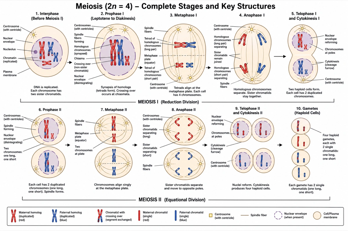
Labeled meiosis diagram (answer key)
Every stage of meiosis I and meiosis II in order, with the stages and key events named. Print it as the answer key, or hand it out as a revision sheet.
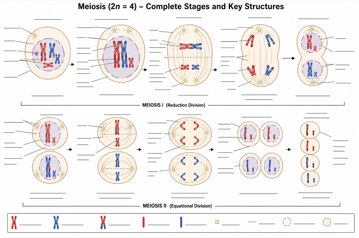
Blank meiosis diagram to label
The same drawing with an empty answer line where every label was, ready for a worksheet, a quiz or an exam question.
What is a meiosis diagram?
A meiosis diagram shows one diploid cell dividing twice into four haploid cells: homologous chromosomes pair, cross over and separate in meiosis I, and sister chromatids separate in meiosis II. Each stage is drawn in order.
Key facts
- One round of DNA replication, two divisions. Chromosomes are copied once, in the S phase before meiosis, and the cell then divides twice, halving the chromosome number: a human cell goes from 46 chromosomes to 23.
- Meiosis I separates homologous chromosomes; meiosis II separates sister chromatids. After meiosis I each cell is already haploid, though every chromosome still has two chromatids; meiosis II splits them, much as mitosis does.
- Crossing over happens in prophase I. Homologous chromosomes pair tightly (synapsis) and non-sister chromatids exchange segments; the crossover points show as chiasmata, at least one per chromosome.
- Independent assortment multiplies the variety. Each homologous pair lines up at random in metaphase I, so a human cell with 23 pairs can produce over 8 million (2²³) chromosome combinations before crossing over is counted.
- The four products are genetically different. Crossing over and independent assortment make every haploid cell unique, unlike the two identical cells mitosis produces.
How does the meiosis diagram maker work?
Describe the diagram
Say what it is for and how deep it should go — every stage fully labeled, a simple overview, meiosis I and meiosis II side by side. A sentence is enough.
Pick a style
The default is a clean flat textbook plate — white background, labels on straight leader lines. Or apply watercolor, ink line art, 3D and other finishes.
Refine and export
The result opens in your workspace, where you can redraw regions, edit labels on the canvas, upscale and export in high resolution.
Stages of meiosis and what happens in each
The stages a labeled meiosis diagram is normally expected to show, what happens to the chromosomes in each, and what to draw. This is the default label set unless you ask for a shorter one.
| Stage | What happens | What to draw |
|---|---|---|
| Interphase (before meiosis) | DNA is replicated; each chromosome becomes two sister chromatids | One diploid cell with an intact nucleus |
| Prophase I | Chromosomes condense; homologous chromosomes pair (synapsis) and cross over | Paired homologs joined at chiasmata, nuclear envelope breaking down |
| Metaphase I | Homologous pairs line up at the metaphase plate, each in random orientation | Pairs side by side across the equator, spindle fibers from both poles |
| Anaphase I | Homologous chromosomes are pulled to opposite poles; sister chromatids stay together | Whole X-shaped chromosomes moving apart |
| Telophase I and cytokinesis | Two haploid cells form, each chromosome still made of two chromatids | Two cells, each with half the chromosomes |
| Prophase II | Chromosomes condense again and new spindles form, with no DNA replication | Two cells, each with a new spindle |
| Metaphase II | Chromosomes line up singly at the plate in each cell | One row of X-shaped chromosomes per cell |
| Anaphase II | Sister chromatids separate and move to opposite poles | Single chromatids moving apart in both cells |
| Telophase II and cytokinesis | Nuclei re-form and both cells divide | Four haploid cells, each genetically different |
Courses differ in how many stages they expect. Introductory worksheets often show meiosis I and II as two steps ending in four cells; advanced courses add prometaphase, interkinesis, the synaptonemal complex and the substages of prophase I. Name the stages you need and only those are drawn.
Mitosis vs meiosis: what is the difference?
Mitosis is one division that makes two cells genetically identical to the parent, with the same chromosome number; meiosis is two divisions that make four genetically different cells with half the chromosome number. Mitosis builds and repairs the body; meiosis makes the haploid cells of sexual reproduction — eggs and sperm in animals.
| Mitosis | Meiosis | |
|---|---|---|
| Number of divisions | One | Two |
| Cells produced | Two | Four |
| Chromosome number | Same as the parent | Halved, diploid to haploid |
| Genetic makeup | Identical to the parent | Different from the parent and from each other |
| Homologous chromosomes pair up | No | Yes, in prophase I |
| Crossing over | No | Yes, in prophase I |
| What lines up at the metaphase plate | Single chromosomes | Homologous pairs in metaphase I, single chromosomes in metaphase II |
| Purpose | Growth, repair and asexual reproduction | Gametes or spores for sexual reproduction |
| Where in the human body | Body (somatic) cells | Ovaries and testes |
Draw meiosis I with pairs of X-shaped chromosomes and meiosis II with single ones, and keep the maternal and paternal colors through every stage so crossing over shows as swapped segments. A diagram that pairs homologs in mitosis, or splits sister chromatids in anaphase I, is the error to check for first.
Where are meiosis diagrams used?
Meiosis diagrams are used mostly in biology teaching — labeling worksheets, genetics lessons and exam revision — and as schematic panels in talks and papers on fertility, chromosome errors and breeding. What changes is the depth: two divisions and four cells for a first course, every stage with crossing over and chiasmata for a university one.
School biology
Labeling worksheets and revision sheets from middle school through GCSE, A-level, AP Biology and IB. Usually the stages in order, plus the same drawing blank for students to fill in.
Genetics lessons
Diagrams that tie meiosis to inheritance: crossing over and independent assortment explaining why siblings differ, and Mendel's laws following from how chromosomes separate.
Exam revision
One-page comparisons of mitosis and meiosis, and of meiosis I and II — which is how exam questions ask about them: identify the stage, say whether the cell is haploid or diploid.
Medical and research figures
Schematic panels on nondisjunction and aneuploidy — how a failed separation in meiosis I or II leads to a trisomy such as Down syndrome — or on meiosis in plant and animal breeding.
Common questions about meiosis diagrams
What should a labeled meiosis diagram include?
The standard set is prophase I with crossing over, metaphase I, anaphase I and telophase I, then prophase II, metaphase II, anaphase II and telophase II, ending in four haploid cells — with the homologous chromosomes, sister chromatids, chiasmata, spindle fibers and metaphase plate labeled. That is the default — ask for fewer labels and only those are drawn.
Can I get a blank meiosis diagram for students to label?
Yes — view the blank version on this page free, or ask for one and every label is replaced by an empty answer line. The labeled version is the matching answer key, so the two print as a pair.
What are the stages of meiosis in order?
Prophase I, metaphase I, anaphase I and telophase I, then prophase II, metaphase II, anaphase II and telophase II. Many textbooks also name prometaphase I and II, and some species pass through a short interkinesis between the two divisions, with no DNA replication.
What is the difference between meiosis I and meiosis II?
Meiosis I separates homologous chromosomes and halves the chromosome number; meiosis II separates sister chromatids and keeps the number the same. Crossing over and independent assortment both happen in meiosis I; meiosis II works much like mitosis in a haploid cell.
What is crossing over, and when does it happen?
Crossing over is the exchange of segments between non-sister chromatids of a homologous pair, during prophase I. A protein lattice, the synaptonemal complex, holds the pair together while the DNA is cut and rejoined; the crossover points appear as chiasmata, which keep the pair linked until anaphase I.
Why does meiosis produce genetic variation?
Because of crossing over in prophase I and the random orientation of homologous pairs in metaphase I. With 23 chromosome pairs, a human can make over 8 million chromosome combinations from independent assortment alone; crossing over multiplies that further.
What is the difference between haploid and diploid?
A diploid cell has two sets of chromosomes, one from each parent; a haploid cell has one set. In humans, body cells are diploid with 46 chromosomes (2n = 46) and eggs and sperm are haploid with 23 (n = 23); fertilization restores the diploid number.
What happens if chromosomes fail to separate in meiosis?
This is nondisjunction, and it gives gametes an extra or a missing chromosome. If it happens in meiosis I, all four gametes are abnormal; in meiosis II, two of the four are. A fertilized egg with an extra chromosome 21 — trisomy 21 — develops Down syndrome.
Can I make a simple meiosis drawing for younger students?
Yes — ask for a simple overview and the diagram drops to the essentials: one parent cell dividing twice into four sex cells, each with half the chromosomes. Five labels or fewer usually covers a middle-school class.
How is this different from a generic AI image tool?
The subject brief is built in. Every render is instructed to follow the standard textbook sequence — each stage drawn in its true order and joined to the next by an arrow — with each stage name spelled exactly. Check the chromosome counts and labels against your course before printing; the canvas lets you correct any label in place.
Can I use the diagrams for school, teaching or publication?
Yes — diagrams you generate are yours to use in worksheets, presentations, handouts, theses and papers. Export high-resolution raster up to 4K depending on the model.
Can I change the labels after generating?
Yes. The result opens in a canvas workspace that recognizes the diagram labels — you can edit or translate the text, move leader lines, redraw a region or recolor without regenerating the whole figure.
Sources
Keep exploring
Our other diagram tools, and the guides behind them.

Draw your meiosis diagram
From a one-line description to a labeled, printable diagram in about a minute.
Start drawing