
Protein Synthesis Diagram Maker
Describe the diagram you need and get a labeled protein synthesis diagram — transcription in the nucleus, mRNA processing, and translation at the ribosome with tRNAs, codons and anticodons, in order. Or view the labeled and blank versions below, free.
Or start from an example
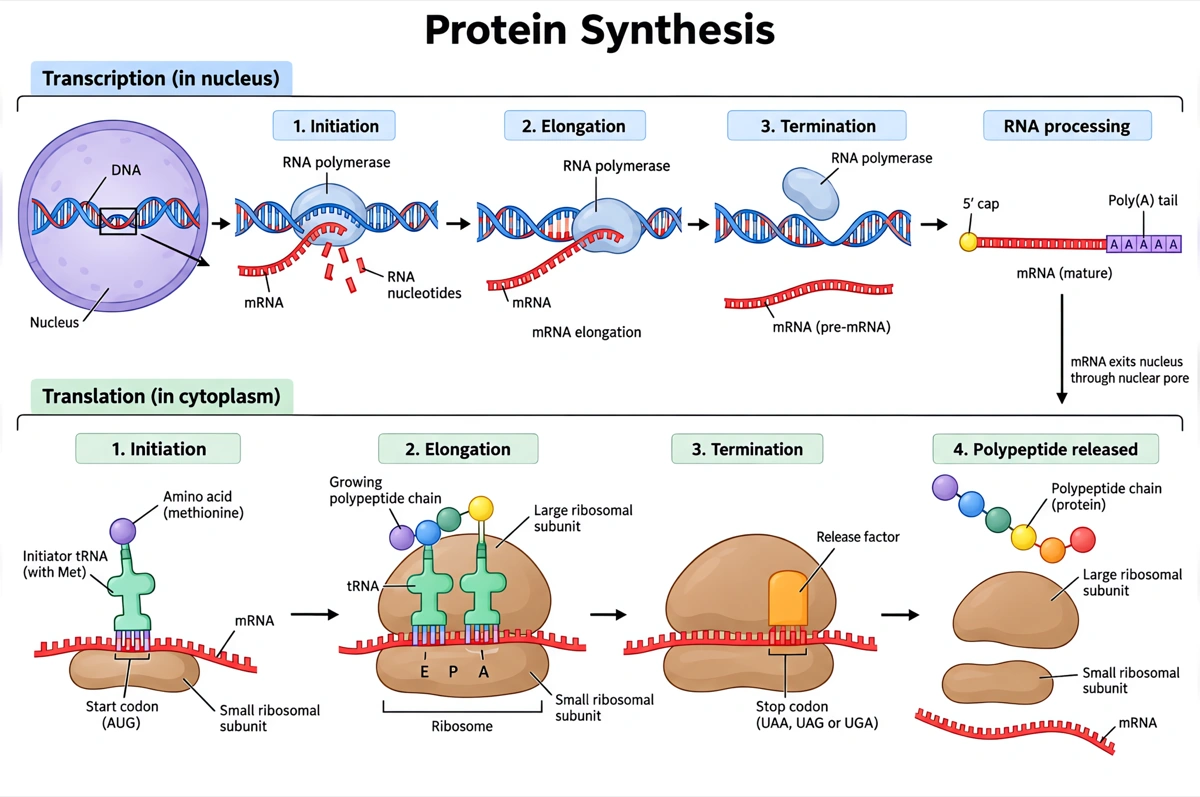
Labeled protein synthesis diagram (answer key)
Transcription and translation in order, with the molecules and the places they happen named. Print it as the answer key, or hand it out as a revision sheet.
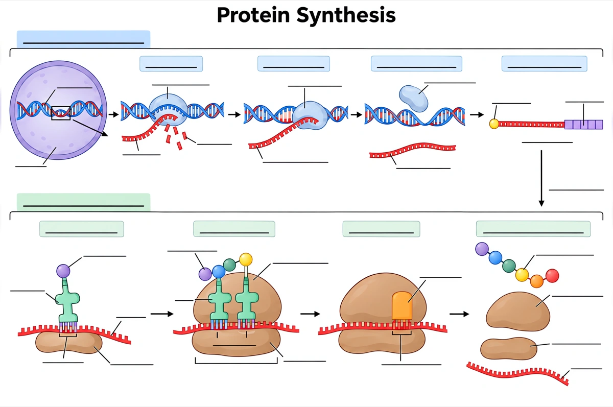
Blank protein synthesis diagram to label
The same drawing with an empty answer line where every label was, ready for a worksheet, a quiz or an exam question.
What is a protein synthesis diagram?
A protein synthesis diagram shows how a cell turns a gene into a protein in two stages: transcription, where RNA polymerase copies DNA into mRNA, and translation, where a ribosome reads its codons and tRNAs bring the matching amino acids. In eukaryotes the mRNA is processed in between.
Key facts
- Information flows from DNA to RNA to protein. This is the central dogma: transcription makes a messenger RNA copy of a gene, and translation on ribosomes turns that message into a chain of amino acids.
- The genetic code is read in triplets. Each three-base codon on the mRNA specifies one amino acid; of the 64 possible codons, 61 code for amino acids and 3 are stop codons, and AUG both codes for methionine and starts translation.
- Eukaryotic mRNA is processed before it leaves the nucleus. The pre-mRNA gets a 5′ cap and a poly-A tail of about 200 adenines, and its introns are spliced out so that only the exons remain.
- tRNAs are the adaptors. Each tRNA carries one amino acid and has an anticodon that base-pairs with a codon on the mRNA; depending on the species, 40 to 60 kinds of tRNA exist in the cytoplasm.
- The ribosome has three tRNA sites. A new tRNA enters the A site, the tRNA holding the growing chain sits in the P site, and empty tRNAs leave from the E site; peptidyl transferase, an RNA-based enzyme in the large subunit, forms each peptide bond.
How does the protein synthesis diagram maker work?
Describe the diagram
Say what it is for and how deep it should go — the full process in a eukaryotic cell, a simple overview, transcription and translation side by side. A sentence is enough.
Pick a style
The default is a clean flat textbook plate — white background, labels on straight leader lines. Or apply watercolor, ink line art, 3D and other finishes.
Refine and export
The result opens in your workspace, where you can redraw regions, edit labels on the canvas, upscale and export in high resolution.
Stages of protein synthesis and what happens in each
The stages a labeled protein synthesis diagram is normally expected to show, what happens in each, and where in the cell it takes place. This is the default label set unless you ask for a shorter one.
| Stage | What happens | Where it happens |
|---|---|---|
| Transcription: initiation | RNA polymerase binds the promoter and the DNA unwinds | Nucleus (cytoplasm in bacteria) |
| Transcription: elongation | RNA polymerase reads the template strand and builds pre-mRNA 5′ to 3′ | Nucleus |
| Transcription: termination | The transcript is released and the DNA rewinds | Nucleus |
| 5′ capping | A modified guanine cap is added to the front of the pre-mRNA, protecting it and helping ribosomes bind | Nucleus |
| Poly-A tail | About 200 adenines are added to the 3′ end | Nucleus |
| Splicing | Introns are cut out and exons joined into the mature mRNA | Nucleus |
| mRNA export | The mature mRNA leaves the nucleus through a nuclear pore | Nuclear envelope |
| Translation: initiation | The small ribosomal subunit binds the mRNA, the initiator tRNA pairs with the AUG start codon, and the large subunit joins | Cytoplasm, on free ribosomes or the rough ER |
| Translation: elongation | tRNAs enter the A site, peptide bonds join the amino acids, and the ribosome moves along the mRNA one codon at a time | Ribosome |
| Translation: termination | A stop codon reaches the A site, a release factor binds, and the polypeptide is released | Ribosome |
Courses differ in how finely they split the process. Introductory worksheets often show two steps, transcription and translation, with DNA, mRNA, the ribosome and tRNA; A-level and university courses add RNA processing, the A, P and E sites, the start and stop codons and the three phases of each stage. Name the labels you need and only those are drawn.
Transcription vs translation: what is the difference?
Transcription copies a gene's DNA into messenger RNA; translation reads that mRNA to build a protein. In eukaryotes transcription happens in the nucleus and translation in the cytoplasm; in bacteria, which have no nucleus, both happen in the cytoplasm.
| Transcription | Translation | |
|---|---|---|
| Template | DNA (the template strand) | mRNA |
| Product | RNA — pre-mRNA in eukaryotes | A polypeptide chain |
| Where, in eukaryotes | Nucleus | Cytoplasm, on ribosomes |
| Main machinery | RNA polymerase (RNA polymerase II for mRNA) | Ribosome and tRNAs |
| Building blocks | RNA nucleotides (A, U, G, C) | Amino acids |
| Start signal | Promoter, such as the TATA box | AUG start codon |
| Stop signal | Termination sequence | Stop codon (UAA, UAG or UGA) |
| Base pairing | RNA nucleotides pair with the DNA template, U in place of T | tRNA anticodons pair with mRNA codons |
Draw transcription inside the nucleus and translation outside it, with the mRNA crossing the nuclear envelope between them, and show at least one codon paired with its tRNA's anticodon. A diagram with the ribosome translating inside the nucleus, or with T in the mRNA, is the error to check for first.
Where are protein synthesis diagrams used?
Protein synthesis diagrams are used mostly in biology teaching — labeling worksheets, genetics lessons and exam revision — and as schematic panels in molecular biology, medicine and biotechnology. What changes is the depth: DNA, mRNA and a ribosome for a first course, RNA processing and the ribosome's A, P and E sites for a university one.
School biology
Labeling worksheets and revision sheets from middle school through GCSE, A-level, AP Biology and IB. Usually the whole process in order, plus the same drawing blank for students to fill in.
Genetics lessons
Diagrams that tie the genetic code to mutations: how one changed base alters a codon, and how inserting one or two bases shifts the reading frame.
Exam revision
One-page summaries of transcription and translation side by side — which is how exam questions ask about them: name the molecule at X, give the anticodon for a codon, say where each stage happens.
Medicine and biotechnology
Panels on antibiotics that target bacterial ribosomes — tetracycline blocks the A site, chloramphenicol blocks peptide bond formation — and on making proteins from engineered genes.
Common questions about protein synthesis diagrams
What should a labeled protein synthesis diagram include?
The standard set is the DNA with its template strand, RNA polymerase and the mRNA in the nucleus, a nuclear pore, then the ribosome with its small and large subunits, tRNAs with their anticodons and amino acids, the mRNA codons from the start codon on, and the growing polypeptide chain. That is the default — ask for fewer labels and only those are drawn.
Can I get a blank protein synthesis diagram for students to label?
Yes — view the blank version on this page free, or ask for one and every label is replaced by an empty answer line. The labeled version is the matching answer key, so the two print as a pair.
What are the steps of protein synthesis in order?
Transcription, RNA processing and translation. RNA polymerase copies a gene into pre-mRNA; in eukaryotes the pre-mRNA is capped, given a poly-A tail and spliced; the mRNA leaves the nucleus; and a ribosome translates it — initiation at AUG, elongation codon by codon, termination at a stop codon.
What is the difference between a codon and an anticodon?
A codon is a three-base sequence on the mRNA; an anticodon is the matching three-base sequence on a tRNA. The two pair by complementary base pairing: the codon AUG is read by a tRNA with the anticodon UAC, which carries methionine.
What do mRNA, tRNA and rRNA do?
mRNA carries the gene's message from the nucleus to the ribosome; tRNA brings the amino acids and matches them to the codons; rRNA forms the core of the ribosome and catalyzes the peptide bonds. All three are made by transcription.
Where does protein synthesis take place?
Transcription takes place in the nucleus and translation in the cytoplasm, on free ribosomes or ribosomes on the rough endoplasmic reticulum. In bacteria, which have no nucleus, both take place in the cytoplasm. Mitochondria and chloroplasts also have ribosomes of their own.
How many codons are there?
64: 61 code for amino acids and 3 are stop codons. Most amino acids have more than one codon, which is why the code is called degenerate; AUG is the start codon and also codes for methionine.
How does a mutation change protein synthesis?
A changed base changes the codon, which may then call for a different amino acid, the same one, or a stop. Inserting or deleting one or two bases shifts the reading frame and garbles every codon after it — the effect Crick and Brenner used to show that the code is read in triplets.
Can I make a simple protein synthesis drawing for younger students?
Yes — ask for a simple overview and the diagram drops to the essentials: DNA in the nucleus, the mRNA copy, the ribosome, and the amino acids joining into a protein. Five labels or fewer usually covers a middle-school class.
How is this different from a generic AI image tool?
The subject brief is built in. Every render is instructed to follow the standard textbook sequence — each stage drawn in its true order and joined to the next by an arrow — with each label spelled exactly. Check the labels against your course before printing; the canvas lets you correct any in place.
Can I use the diagrams for school, teaching or publication?
Yes — diagrams you generate are yours to use in worksheets, presentations, handouts, theses and papers. Export high-resolution raster up to 4K depending on the model.
Can I change the labels after generating?
Yes. The result opens in a canvas workspace that recognizes the diagram labels — you can edit or translate the text, move leader lines, redraw a region or recolor without regenerating the whole figure.
Sources
Keep exploring
Our other diagram tools, and the guides behind them.

Draw your protein synthesis diagram
From a one-line description to a labeled, printable diagram in about a minute.
Start drawing