
Nephron Diagram Maker
Describe the diagram you need and get a labeled nephron — glomerulus, Bowman's capsule, proximal and distal convoluted tubules, loop of Henle and collecting duct, with the blood vessels that serve them. Or view the labeled and blank versions below, free.
Or start from an example
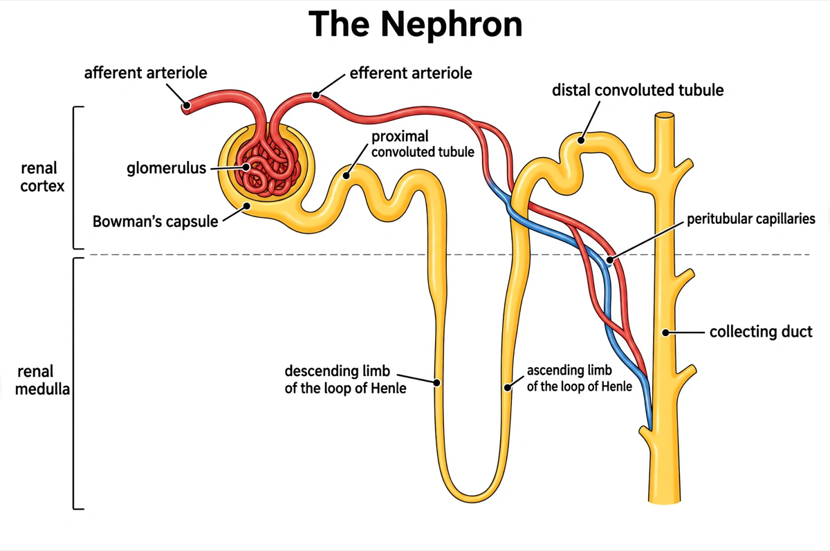
Labeled nephron diagram (answer key)
Every part of the nephron and its blood supply named, from the glomerulus to the collecting duct. Print it as the answer key, or hand it out as a revision sheet.
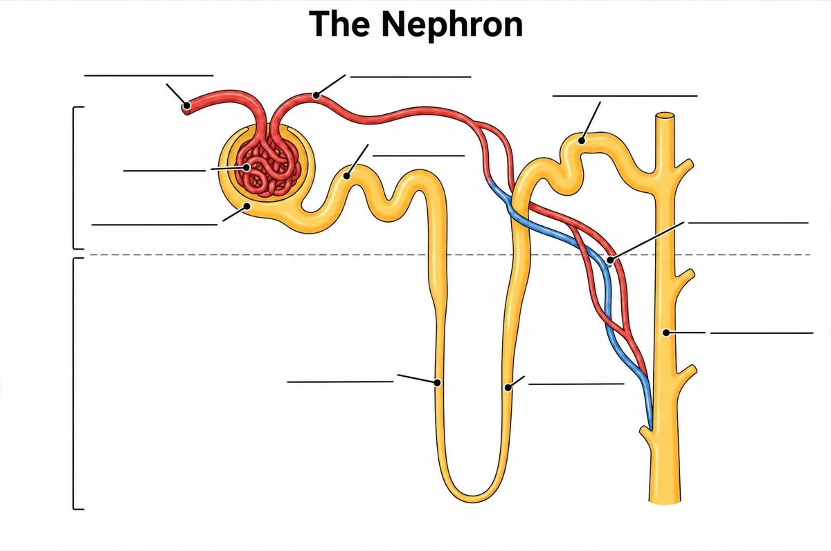
Blank nephron diagram to label
The same drawing with an empty answer line where every label was, ready for a worksheet, a quiz or an exam question.
What is a nephron diagram?
A nephron diagram shows the kidney's filtering unit: the glomerulus filters blood into Bowman's capsule, and the filtrate flows through the proximal convoluted tubule, the loop of Henle and the distal convoluted tubule into a collecting duct. Each part is labeled with its job.
Key facts
- Each kidney holds over a million nephrons. About 85 percent are cortical nephrons, in the renal cortex with short loops of Henle; about 15 percent are juxtamedullary nephrons, next to the medulla, whose long loops reach deep into it.
- A nephron does three jobs: filtration, reabsorption and secretion. The glomerulus filters the blood, the tubules take back what the body needs, and wastes such as urea, creatinine and ammonia, excess K⁺ and some drugs are secreted into the filtrate.
- Up to 180 liters of filtrate a day become 1 to 2 liters of urine. Both kidneys together filter about 125 mL of plasma a minute in men and 105 mL in women, and about 99 percent of that filtrate is returned to the blood.
- The proximal convoluted tubule does most of the reclaiming. It reabsorbs about 65 percent of the sodium and 67 percent of the water in the filtrate and almost all of the glucose and amino acids, across a brush border of microvilli.
- The loop of Henle lets the kidney concentrate urine. Water leaves its descending limb and salt is pumped out of its ascending limb, building a salt gradient in the medulla that the collecting duct uses, under ADH, to take back water.
How does the nephron diagram maker work?
Describe the diagram
Say what it is for and how deep it should go — a full labeled nephron with its blood vessels, a simple overview, cortical and juxtamedullary nephrons side by side. A sentence is enough.
Pick a style
The default is a clean flat textbook plate — white background, labels on straight leader lines. Or apply watercolor, ink line art, 3D and other finishes.
Refine and export
The result opens in your workspace, where you can redraw regions, edit labels on the canvas, upscale and export in high resolution.
Parts of the nephron and what each one does
The parts a labeled nephron diagram is normally expected to show, what each one does, and where it sits in the kidney. This is the default label set unless you ask for a shorter one.
| Part | What it does | Where it sits |
|---|---|---|
| Afferent arteriole | Brings blood into the glomerulus | Renal cortex, entering the renal corpuscle |
| Glomerulus | A tuft of high-pressure capillaries that filters water, salts, glucose and urea out of the blood, holding back cells and large proteins | Renal cortex, inside Bowman's capsule |
| Efferent arteriole | Carries blood out of the glomerulus to the peritubular capillaries | Renal cortex, leaving the renal corpuscle |
| Bowman's capsule | Cup that catches the filtrate and funnels it into the tubule | Renal cortex, around the glomerulus |
| Proximal convoluted tubule (PCT) | Reabsorbs most of the water and sodium and almost all of the glucose and amino acids | Renal cortex |
| Descending limb of the loop of Henle | Lets water leave the filtrate by osmosis | Dips into the renal medulla |
| Ascending limb of the loop of Henle | Moves Na⁺ and Cl⁻ out but keeps water in, diluting the filtrate | Rises from the medulla back to the cortex |
| Distal convoluted tubule (DCT) | Secretes K⁺ and H⁺ and reabsorbs Na⁺, Cl⁻ and HCO₃⁻, fine-tuning salt balance and pH | Renal cortex |
| Collecting duct | Gathers filtrate from several nephrons and reabsorbs water when ADH is present | From the cortex down through the medulla |
| Peritubular capillaries and vasa recta | Carry the reabsorbed water and solutes back into the blood | Around the tubules; the vasa recta beside the loop of Henle |
Courses differ in how many parts they expect. Introductory worksheets often stop at the glomerulus, Bowman's capsule, the tubule, the loop of Henle and the collecting duct; A-level and university courses split the tubule into PCT and DCT and add the afferent and efferent arterioles, the vasa recta and the juxtaglomerular apparatus. Name the labels you need and only those are drawn.
Cortical vs juxtamedullary nephron: what is the difference?
Cortical nephrons lie in the renal cortex and have short loops of Henle that barely reach the medulla; juxtamedullary nephrons lie next to the medulla and have long loops that run deep into it. About 85 percent of nephrons are cortical; the 15 percent that are juxtamedullary build the salt gradient that concentrates urine.
| Cortical nephron | Juxtamedullary nephron | |
|---|---|---|
| Share of nephrons | About 85 percent | About 15 percent |
| Renal corpuscle | In the renal cortex, away from the medulla | In the cortex, next to the medulla |
| Loop of Henle | Short, entering the medulla little if at all | Long, some reaching deep into the medulla |
| Capillaries around the tubule | Peritubular capillaries around the PCT and DCT | Peritubular capillaries plus the vasa recta along the loop |
| Main role | Everyday filtration and reabsorption | Building the salt gradient that concentrates urine |
Draw the juxtamedullary loop hairpinning deep into the medulla with the vasa recta beside it, and the cortical loop staying short near the cortex. A diagram with both loops the same length, or with the collecting duct drawn as one nephron's own tubule rather than shared by several, is the error to check for first.
Where are nephron diagrams used?
Nephron diagrams are used mostly in biology and physiology teaching — labeling worksheets, exam revision and lecture slides — and in medical, nursing and pharmacology courses that show where hormones and drugs act along the tubule. What changes is the depth: five parts for a first course, every segment with what it moves for a medical one.
School biology
Labeling worksheets and revision sheets for GCSE, A-level, AP Biology and IB, where the nephron is the core of excretion and osmoregulation. Usually the labeled nephron, plus the same drawing blank for students to fill in.
Medicine and nursing
Physiology and pharmacology slides that put each control on its segment: ADH and aldosterone on the collecting duct, loop diuretics on the ascending limb of the loop of Henle.
Exam revision
One-page summaries that match each part to its job — which is how exam questions ask about them: name the structure at X, explain why glucose appears in the urine in untreated diabetes.
Research and patient education
Schematic panels for kidney research and patient leaflets on kidney disease, with the affected part of the nephron highlighted.
Common questions about nephron diagrams
What should a labeled nephron diagram include?
The standard set is the afferent arteriole, glomerulus, efferent arteriole, Bowman's capsule, proximal convoluted tubule, descending and ascending limbs of the loop of Henle, distal convoluted tubule and collecting duct, often with the peritubular capillaries and the renal cortex and medulla marked. That is the default — ask for fewer labels and only those are drawn.
Can I get a blank nephron diagram for students to label?
Yes — view the blank version on this page free, or ask for one and every label is replaced by an empty answer line. The labeled version is the matching answer key, so the two print as a pair.
What are the parts of a nephron in order?
The glomerulus and Bowman's capsule (together the renal corpuscle), then the proximal convoluted tubule, the descending limb of the loop of Henle, the ascending limb and the distal convoluted tubule, which empties into a collecting duct. The collecting duct serves several nephrons and is continuous with the nephron but not strictly part of it.
What is the function of the nephron?
It filters the blood and turns the filtrate into urine, in three steps: glomerular filtration, tubular reabsorption and tubular secretion. Along the way it keeps the blood's water, salt and pH in balance, and the nephron's juxtaglomerular cells help control blood pressure by releasing renin.
What does the loop of Henle do?
It builds a salt gradient in the renal medulla that lets the kidney make concentrated urine. Water leaves the descending limb by osmosis; salt is moved out of the ascending limb, which lets no water follow. Because the two limbs work in opposite ways, the loop acts as a countercurrent multiplier.
Where is most water reabsorbed in the nephron?
In the proximal convoluted tubule, which takes back about two-thirds of the water in the filtrate. The descending limb of the loop of Henle reclaims about 15 percent more and the distal convoluted tubule about 8 percent; the collecting duct takes back a variable amount set by ADH — almost all that reaches it when you are dehydrated, almost none when you are over-hydrated.
What does ADH do in the nephron?
ADH (antidiuretic hormone) makes the collecting duct permeable to water. It signals the duct's cells to insert aquaporin water channels into their membranes, so water leaves the filtrate and returns to the blood and the urine is more concentrated. Without ADH the channels are not inserted and dilute urine is excreted.
Why is there no glucose in normal urine?
Because the proximal convoluted tubule reabsorbs almost 100 percent of the filtered glucose. Glucose passes freely into the filtrate and is carried back into the blood together with sodium. It appears in the urine only when blood glucose is high enough to saturate those carriers — usually a sign of type 1 or type 2 diabetes.
Can I make a simple nephron drawing for younger students?
Yes — ask for a simple overview and the diagram drops to the essentials: the glomerulus, Bowman's capsule, the tubule, the loop of Henle and the collecting duct. Five labels or fewer usually covers a first biology class.
How is this different from a generic AI image tool?
The subject brief is built in. Every render is instructed to follow the standard textbook drawing — every part of the nephron in its true position, order and relative proportion — with each label spelled exactly and set on a straight leader line. Check the labels against your course before printing; the canvas lets you correct any in place.
Can I use the diagrams for school, teaching or publication?
Yes — diagrams you generate are yours to use in worksheets, presentations, handouts, theses and papers. Export high-resolution raster up to 4K depending on the model.
Can I change the labels after generating?
Yes. The result opens in a canvas workspace that recognizes the diagram labels — you can edit or translate the text, move leader lines, redraw a region or recolor without regenerating the whole figure.
Keep exploring
Our other diagram tools, and the guides behind them.

Draw your nephron diagram
From a one-line description to a labeled, printable diagram in about a minute.
Start drawing