
Neuron Diagram Maker
Describe the diagram you need and get a labeled neuron — dendrites, cell body and nucleus, axon hillock, the axon with its myelin sheath and nodes of Ranvier, and the axon terminals. Or view the labeled and blank versions below, free.
Neuron diagram examples
Real renders of the prompts shown: a fully labeled motor neuron, a six-label neuron drawing for beginners, sensory, inter- and motor neurons side by side and a blank worksheet. Click one to load and edit its prompt.
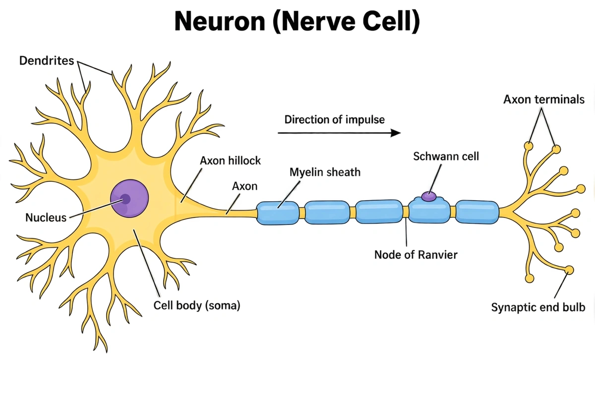
Labeled neuron diagram (answer key)
Every part of a motor neuron named, from the dendrites to the axon terminals, with the myelin sheath, the nodes of Ranvier and a Schwann cell. Print it as the answer key, or hand it out as a revision sheet.
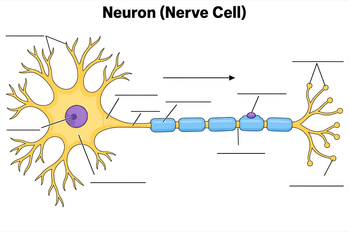
Blank neuron diagram to label
The same drawing with an empty answer line where every label was, ready for a worksheet, a labeling quiz or an exam question.
What is a neuron diagram?
A neuron diagram shows the parts of a nerve cell in the order a signal passes through them: the dendrites receive it, the cell body integrates it, and the axon carries it from the axon hillock, past the myelin sheath and nodes of Ranvier, to the axon terminals. Most textbooks draw a multipolar motor neuron, labeled part by part with the impulse running left to right.
Key facts
- A signal runs one way through a neuron: dendrites → cell body → axon → axon terminals. The dendrites and cell body collect inputs from other cells, and only the axon carries the impulse on to the next neuron, muscle fiber or gland.
- The action potential starts at the axon hillock. This trigger zone, where the cell body tapers into the axon, fires once the summed inputs lift the membrane from its resting −70 mV to a threshold of about −55 mV.
- Myelin raises conduction speed from about 0.5–2 m/s to as much as 120 m/s. The impulse jumps from one node of Ranvier to the next — saltatory conduction — because the voltage-gated Na⁺ channels are concentrated in those bare gaps, about 1 mm apart.
- Schwann cells make the myelin in the peripheral nervous system; oligodendrocytes make it in the brain and spinal cord. Each Schwann cell wraps one segment of a single axon, while one oligodendrocyte can myelinate segments of many axons.
- Neurons are sorted three ways by job: sensory neurons, interneurons and motor neurons. Sensory neurons bring signals in from receptors, interneurons connect neurons inside the brain and spinal cord, and motor neurons carry commands out to muscles and glands; by shape, neurons are multipolar, bipolar or unipolar.
How does the neuron diagram maker work?
Describe the diagram
Say what it is for and how deep it should go — a full labeled motor neuron, a simple drawing for beginners, sensory and motor neurons side by side. A sentence is enough.
Pick a style
The default is a clean flat textbook plate — white background, labels on straight leader lines. Or apply watercolor, ink line art, 3D and other finishes.
Refine and export
The result opens in your workspace, where you can redraw regions, edit labels on the canvas, upscale and export in high resolution.
Parts of a neuron and what each one does
The parts a labeled neuron diagram is normally expected to show, what each one does, and where it sits along the cell, in the order a signal passes through them. This is the default label set unless you ask for a shorter one.
| Part | What it does | Where it sits |
|---|---|---|
| Dendrites | Receive signals from other neurons and pass them toward the cell body | Branching out from the cell body; the start of the signal path |
| Cell body (soma) | Integrates the incoming signals and keeps the cell alive, making its proteins | Between the dendrites and the axon |
| Nucleus | Holds the cell's DNA and directs protein synthesis | Inside the cell body |
| Axon hillock | Sums the inputs and starts the action potential when the membrane reaches threshold | Where the cell body tapers into the axon |
| Axon | Carries the action potential away from the cell body — about 1 m long in the motor neurons that reach the foot | From the axon hillock to the axon terminals |
| Myelin sheath | Insulates the axon so the impulse jumps between gaps and travels much faster | Wrapped around the axon in segments |
| Schwann cell | Forms one myelin segment around a peripheral axon and helps a cut peripheral axon regrow | Along the axon, with its nucleus on the outside of the sheath |
| Node of Ranvier | A bare gap packed with voltage-gated Na⁺ channels, where the impulse is regenerated | Between neighboring myelin segments, about 1 mm apart |
| Axon terminals | Release neurotransmitter from synaptic vesicles to pass the signal on | The branched far end of the axon, each ending in a synaptic end bulb |
| Synapse | The junction where the signal crosses to the next cell, over a cleft about 20–40 nm wide | Between an axon terminal and a dendrite, muscle fiber or gland cell |
Courses differ in how many parts they expect. Introductory worksheets often stop at the dendrites, cell body, nucleus, axon and axon terminals; GCSE, A-level and anatomy-and-physiology courses add the myelin sheath, nodes of Ranvier and Schwann cells, and university courses the axon hillock, Nissl bodies and synaptic end bulbs. Name the labels you need and only those are drawn.
Sensory vs motor neuron: what is the difference?
Sensory neurons carry signals from receptors into the brain and spinal cord; motor neurons carry commands out of them to muscles and glands. They also look different: a sensory neuron is usually unipolar, with its cell body to one side of the axon, while a motor neuron is multipolar, with its dendrites around the cell body at the start of the axon.
| Sensory neuron | Motor neuron | |
|---|---|---|
| Also called | Afferent neuron | Efferent neuron |
| Direction of the signal | From a receptor toward the brain and spinal cord | From the brain and spinal cord to a muscle or gland |
| Shape | Usually unipolar (pseudounipolar): one process that splits into a peripheral and a central branch | Multipolar: many dendrites and one long axon |
| Cell body | To the side of the axon, in a ganglion such as the dorsal root ganglion | At the start of the axon, in the spinal cord's ventral horn or the brainstem |
| Dendrites | At the receptor end, far from the cell body | Branching straight from the cell body |
| Ends on | Interneurons or motor neurons in the brain and spinal cord | Muscle fibers at the neuromuscular junction, or gland cells |
Check where the cell body sits. A sensory neuron's cell body hangs to one side of the axon on a short stalk, while a motor neuron's sits at the start of the axon among its dendrites; a diagram that draws both the same way, or points the impulse arrow back toward the receptor, is the error to look for first.
Where are neuron diagrams used?
Neuron diagrams are used mostly in biology and anatomy-and-physiology teaching — labeling worksheets, quizzes and lecture slides — and in medicine and neuroscience, where they show which part of the cell a disease or a drug acts on. What changes is the depth: five parts for a first course, the trigger zone and myelin for A&P, the ion channels at each node for medicine.
School biology
Labeling worksheets and revision sheets for GCSE, A-level, AP Biology and IB, where the neuron opens the topic of nervous coordination and the reflex arc. Usually the labeled neuron, plus the same drawing blank for students to fill in.
Anatomy & physiology and nursing
A&P lab sheets and nursing lectures that pair the parts of a neuron with nervous tissue, the glial cells and the reflex arc — the figure students label before they learn how the impulse travels.
Medicine
Neurology and pharmacology slides that mark where a condition strikes: lost myelin in multiple sclerosis, the peripheral nerves in Guillain–Barré syndrome, the Na⁺ channels that local anesthetics block.
Research and textbook figures
Schematic panels for neuroscience papers, posters and textbook chapters — a neuron with the recorded region, the labeled cell type or the injected site highlighted.
Common questions about neuron diagrams
What are the parts of a neuron in order?
In the order a signal travels: dendrites, the cell body (soma) with its nucleus, the axon hillock, the axon — wrapped in a myelin sheath broken by nodes of Ranvier — and the axon terminals. The terminals end at synapses with the next neuron, a muscle fiber or a gland cell.
What should a labeled neuron diagram include?
The standard set is the dendrites, cell body (soma), nucleus, axon hillock, axon, myelin sheath, Schwann cells, nodes of Ranvier and axon terminals, often with an arrow showing the direction of the impulse. That is the default — ask for fewer labels and only those are drawn.
Can I get a blank neuron diagram to label?
Yes — view the blank version on this page free, or ask for one and every label is replaced by an empty answer line. The labeled version is the matching answer key, so the two print as a pair and work as a neuron labeling quiz.
How do you draw a neuron?
Draw a round cell body with its nucleus, add short branching dendrites around it, then run one long axon out from a cone-shaped axon hillock and end it in a few branched axon terminals. Wrap the axon in a row of sausage-shaped myelin segments with small gaps between them for the nodes of Ranvier, then add the labels — or describe the drawing here and it is made for you.
What does the myelin sheath do?
It insulates the axon, so the impulse jumps from one node of Ranvier to the next instead of creeping along the whole membrane — saltatory conduction. That raises the speed from about 0.5–2 m/s in unmyelinated axons to as much as 120 m/s. When myelin is lost, as in multiple sclerosis, the signal slows or fails.
What are the 3 types of neurons?
Sensory neurons, interneurons and motor neurons. Sensory neurons carry signals from receptors into the brain and spinal cord, interneurons connect neurons inside them, and motor neurons carry commands out to muscles and glands. By shape, neurons are also grouped as multipolar, bipolar or unipolar.
What does the axon hillock do?
It adds up the excitatory and inhibitory inputs reaching the cell body and fires an action potential when the membrane reaches threshold, about −55 mV. Together with the initial segment just beyond it, it forms the trigger zone, where voltage-gated Na⁺ channels are dense enough to start the impulse.
What are the nodes of Ranvier?
Short gaps in the myelin sheath, about 1 mm apart, where the axon membrane is bare. Voltage-gated Na⁺ channels cluster there, so the action potential is regenerated at each node and skips the insulated stretch between them.
What is the difference between a dendrite and an axon?
Dendrites carry signals toward the cell body; the axon carries them away from it. A neuron usually has many short, branching dendrites but only one axon, which can be about a meter long and ends in terminals that release neurotransmitter.
How is this different from a generic AI image tool?
The subject brief is built in. Every render is instructed to follow the standard textbook drawing — every part of the neuron in its true position, order and relative proportion — with each label spelled exactly and set on a straight leader line. Check the labels against your course before printing; the canvas lets you correct any in place.
Can I use the diagrams for school, teaching or publication?
Yes — neuron diagrams you generate are yours to use in worksheets, presentations, handouts, theses and papers. Export high-resolution raster up to 4K depending on the model.
Can I change the labels after generating?
Yes. The result opens in a canvas workspace that recognizes the diagram labels — you can edit or translate the text, move leader lines, redraw a region or recolor without regenerating the whole figure.
Sources
Keep exploring
Our other diagram tools, and the guides behind them.
Tools


Draw your neuron diagram
From a one-line description to a labeled, printable diagram in about a minute.
Start drawing