
Skeletal System Diagram Maker
Describe the diagram you need and get a labeled human skeleton — skull, spine, rib cage, pelvis and every major limb bone, each named with its anatomical term. Or view the labeled and blank versions below, free.
Or start from an example
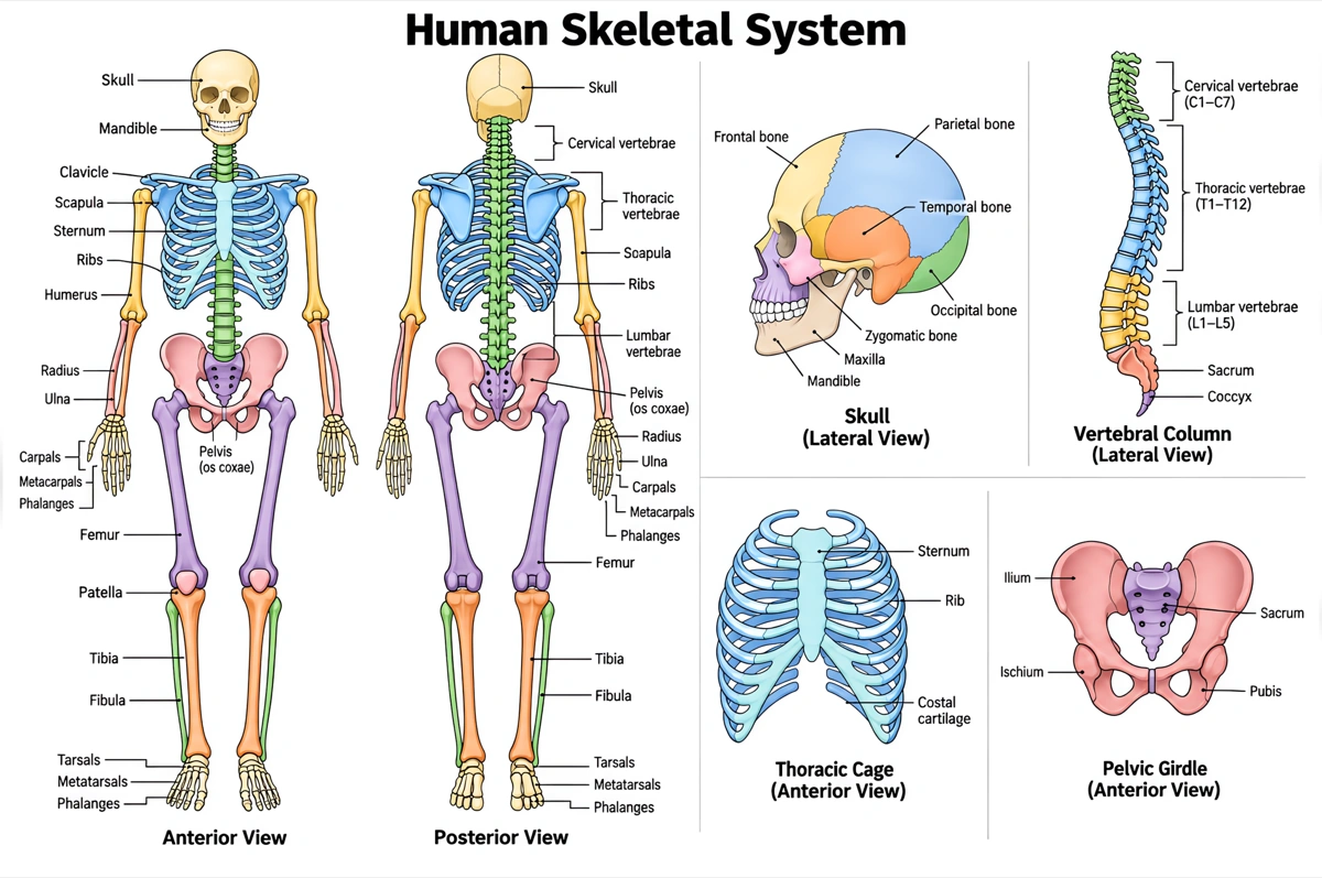
Labeled skeletal system diagram (answer key)
The human skeleton in front view with the major bones named. Print it as the answer key, or hand it out as a revision sheet.
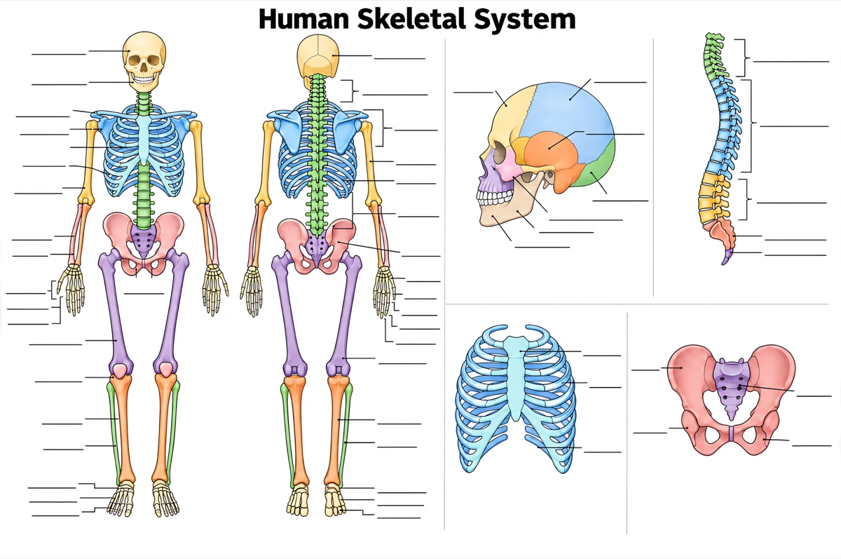
Blank skeletal system diagram to label
The same drawing with an empty answer line where every label was, ready for a worksheet, a quiz or an exam question.
What is a skeletal system diagram?
A skeletal system diagram shows the bones of the human body in place, usually the whole skeleton in front view with the skull, spine, rib cage, pelvis and limb bones labeled. An adult has 206 bones.
Key facts
- An adult has 206 bones; a newborn has more. A baby is born with roughly 270 to 300 bones, some made entirely of cartilage, and many of them fuse during childhood and adolescence.
- The skeleton has two divisions. The axial skeleton — 80 bones of the skull, vertebral column and thoracic cage — forms the body's central axis; the appendicular skeleton — 126 bones — is the limbs plus the shoulder and pelvic girdles that attach them.
- The skeleton does more than hold the body up. It supports the body, gives the muscles levers to move, protects organs such as the brain, heart and lungs, makes blood cells in the red bone marrow, and stores calcium, phosphorus and fat.
- Bones are sorted into five shapes. Long bones (femur, humerus), short bones (carpals, tarsals), flat bones (skull, sternum, ribs), irregular bones (vertebrae) and sesamoid bones (the patella).
- The femur is the longest and strongest bone. The thigh bone accounts for about a quarter of a person's height; each arm and each leg holds 30 bones.
How does the skeletal system diagram maker work?
Describe the diagram
Say what it is for and how deep it should go — a fully labeled skeleton, a simple five-bone version, the axial and appendicular divisions in two colors. A sentence is enough.
Pick a style
The default is a clean flat textbook plate — white background, labels on straight leader lines. Or apply watercolor, ink line art, 3D and other finishes.
Refine and export
The result opens in your workspace, where you can redraw regions, edit labels on the canvas, upscale and export in high resolution.
Major bones of the skeleton and what each one does
The bones a labeled skeletal system diagram is normally expected to name, where each sits, and what it does. This is the default label set unless you ask for a shorter one.
| Bone or group | Where it is | What it does |
|---|---|---|
| Skull (22 bones, including the mandible) | Head | Encloses the brain and shapes the face; the mandible is the only skull bone that moves |
| Vertebral column (24 vertebrae, sacrum and coccyx) | Back, from skull to pelvis | Supports the head and trunk and protects the spinal cord |
| Rib cage (12 pairs of ribs and the sternum) | Chest | Protects the heart and lungs and moves with breathing |
| Clavicle and scapula | Shoulder (pectoral girdle) | Attach each arm to the trunk |
| Humerus | Upper arm | Runs from the shoulder joint to the elbow |
| Radius and ulna | Forearm | Radius on the thumb side, ulna on the little-finger side |
| Carpals, metacarpals and phalanges | Wrist, palm and fingers | 8 carpals, 5 metacarpals and 14 phalanges in each hand |
| Hip bones (pelvic girdle) | Hips | Attach the legs to the vertebral column and carry the trunk's weight |
| Femur | Thigh | Longest and strongest bone, from the hip joint to the knee |
| Patella | Front of the knee | Kneecap, a sesamoid bone inside the thigh muscle's tendon |
| Tibia and fibula | Lower leg | Tibia on the inner side bears the weight; fibula is the thin outer bone |
| Tarsals, metatarsals and phalanges | Ankle, foot and toes | 7 tarsals, 5 metatarsals and 14 phalanges in each foot |
Courses differ in how many bones they expect. Introductory worksheets often stop at the skull, rib cage, spine, pelvis and femur; anatomy courses add both girdles, every long bone and the hand and foot groups, sometimes each carpal by name. Name the labels you need and only those are drawn.
Axial vs appendicular skeleton: what is the difference?
The axial skeleton is the 80 bones along the body's central axis — skull, vertebral column and thoracic cage — and mainly protects the brain, spinal cord, heart and lungs; the appendicular skeleton is the 126 bones of the arms and legs plus the girdles that attach them, and mainly moves the body.
| Axial skeleton | Appendicular skeleton | |
|---|---|---|
| Number of bones (adult) | 80 | 126 |
| Main parts | Skull, hyoid, middle-ear ossicles, vertebral column, thoracic cage | Shoulder girdles, arms, pelvic girdle, legs |
| Position | Along the body's vertical midline | Hanging from the axis on either side |
| Main role | Protection and support of the head and trunk | Movement, walking and handling objects |
| Protects | Brain, spinal cord, heart and lungs | Mainly movement; the pelvic girdle helps shelter the pelvic organs |
| Example bones | Skull, vertebrae, ribs, sternum, sacrum | Clavicle, scapula, humerus, hip bone, femur, tibia |
When the two divisions are colored differently, check the girdles first: the clavicles, scapulae and hip bones belong to the appendicular skeleton even though they sit on the trunk, while the sacrum between the hip bones is axial. A diagram that colors the whole pelvis with the spine has the boundary wrong.
Where are skeletal system diagrams used?
Skeletal system diagrams are used mostly in biology and anatomy teaching — labeling worksheets, exam revision and lecture slides — and in nursing, physiotherapy and sports-science courses that need every bone named correctly. What changes is the depth: five bones for a first course, every long bone and the hand and foot groups for an anatomy one.
School biology
Labeling worksheets and revision sheets from middle school through GCSE, AP Biology and IB. Usually the labeled front-view skeleton, plus the same drawing blank for students to fill in.
Anatomy and health courses
Nursing, physiotherapy, kinesiology and pre-med courses that expect every long bone, both girdles and the carpal and tarsal groups, often split into axial and appendicular.
Exam revision
One-page summaries that put each bone in place — which is how practical exams ask about them: name the bone at X, say whether it is axial or appendicular.
Patient and sports education
Handouts that show where an injury sits — a fractured radius, a stress fracture in the tibia — with the bone highlighted on the whole skeleton.
Common questions about skeletal system diagrams
What should a labeled skeletal system diagram include?
The standard set is the skull, mandible, clavicle, scapula, sternum, ribs, vertebral column, humerus, radius, ulna, carpals, metacarpals, phalanges, pelvis, sacrum, femur, patella, tibia, fibula, tarsals and metatarsals. That is the default — ask for fewer labels and only those are drawn.
Can I get a blank skeletal system diagram for students to label?
Yes — view the blank version on this page free, or ask for one and every label is replaced by an empty answer line. The labeled version is the matching answer key, so the two print as a pair.
How many bones are in the human body?
An adult skeleton has 206 bones: 80 in the axial skeleton and 126 in the appendicular skeleton. A newborn has roughly 270 to 300, some still made of cartilage; many fuse as the child grows, which is why the adult count is lower.
What are the functions of the skeletal system?
Support, movement, protection, blood cell production and storage. The skeleton holds the body up against gravity, gives the muscles levers to pull on, shields the brain, heart and lungs, makes blood cells in the red bone marrow, and stores calcium, phosphorus and fat.
What is the difference between the axial and appendicular skeleton?
The axial skeleton is the 80 bones of the skull, vertebral column and thoracic cage along the body's midline; the appendicular skeleton is the 126 bones of the limbs and the girdles that attach them. The first mainly protects and supports; the second mainly moves.
What is the longest bone in the body?
The femur, or thigh bone — the longest and strongest bone in the body, about a quarter of a person's height. Its rounded head fits into the hip bone to form the hip joint, and its lower end meets the tibia and the patella at the knee.
How many bones are in the hand and the foot?
Each hand has 27 bones — 8 carpals, 5 metacarpals and 14 phalanges — and each foot has 26: 7 tarsals, 5 metatarsals and 14 phalanges. On a whole-skeleton diagram these are usually labeled as groups rather than bone by bone.
What are the five types of bones?
Long, short, flat, irregular and sesamoid. Long bones such as the femur act as levers; short bones (carpals, tarsals) give stability; flat bones (skull, sternum, ribs) protect organs; irregular bones (vertebrae) have complex shapes; sesamoid bones, like the patella, form inside tendons.
Can I make a simple skeleton drawing for younger students?
Yes — ask for a simple overview and the diagram drops to the essentials: the skull, rib cage, spine, pelvis and femur. Five labels or fewer usually covers a middle-school class.
How is this different from a generic AI image tool?
The subject brief is built in. Every render is instructed to follow the standard textbook drawing — every bone in its true position and relative proportion — with each label spelled exactly and set on a straight leader line. Check the labels against your course before printing; the canvas lets you correct any in place.
Can I use the diagrams for school, teaching or publication?
Yes — diagrams you generate are yours to use in worksheets, presentations, handouts, theses and papers. Export high-resolution raster up to 4K depending on the model.
Can I change the labels after generating?
Yes. The result opens in a canvas workspace that recognizes the diagram labels — you can edit or translate the text, move leader lines, redraw a region or recolor without regenerating the whole figure.
Keep exploring
Our other diagram tools, and the guides behind them.

Draw your skeletal system diagram
From a one-line description to a labeled, printable diagram in about a minute.
Start drawing