
Brachial Plexus Diagram Maker
Makes labeled brachial plexus schematics from roots to branches, simple five nerve versions, Erb's and Klumpke's palsy maps and views of the plexus in the neck and axilla. Built for medical, anatomy and anaesthesia students.
Brachial plexus examples
What each prompt drew: the full ladder with all eleven collateral nerves and the posterior divisions in green, a simple version with only the cords and the five terminal nerves, Erb's and Klumpke's palsy shaded over the roots and trunks they hit, and the plexus in place between the scalenes and around the axillary artery with the four block levels numbered. Click a card to load its prompt and change it.
Labeled brachial plexus diagram: every root, trunk, cord and branch
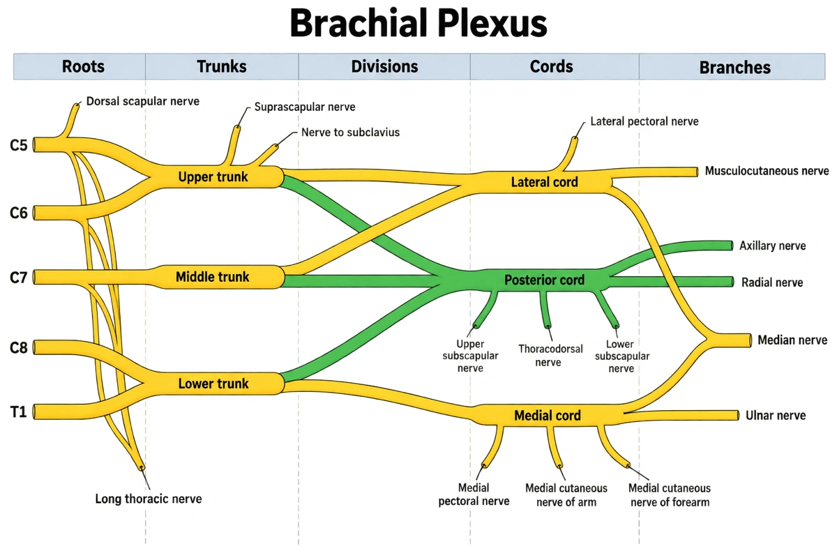
Every labeled part of the ladder, read from the spine outward: where it arises, its root values and what it supplies.
| Part | Arises from | Roots | Supplies |
|---|---|---|---|
| Roots | Anterior rami of the spinal nerves | C5, C6, C7, C8, T1 | Pass between the anterior and middle scalenes to form the trunks |
| Dorsal scapular nerve | C5 root | C5 | Rhomboid major and minor, part of levator scapulae |
| Long thoracic nerve | C5, C6 and C7 roots | C5–C7 | Serratus anterior |
| Upper trunk | C5 and C6 roots | C5–C6 | Anterior and posterior divisions; suprascapular nerve and nerve to subclavius |
| Middle trunk | C7 root alone | C7 | Anterior and posterior divisions; no branches |
| Lower trunk | C8 and T1 roots | C8–T1 | Anterior and posterior divisions; no branches |
| Suprascapular nerve | Upper trunk | C5–C6 | Supraspinatus, infraspinatus, shoulder joint |
| Nerve to subclavius | Upper trunk | C5–C6 | Subclavius |
| Anterior divisions | Front of each trunk | C5–T1 | Lateral and medial cords, the flexor side of the limb |
| Posterior divisions | Back of each trunk | C5–T1 | Posterior cord, the extensor side of the limb |
| Lateral cord | Anterior divisions of upper and middle trunks | C5–C7 | Lateral pectoral, musculocutaneous, lateral root of median |
| Posterior cord | Posterior divisions of all three trunks | C5–T1 | Subscapular and thoracodorsal nerves, axillary, radial |
| Medial cord | Anterior division of lower trunk | C8–T1 | Medial pectoral, medial cutaneous nerves, medial root of median, ulnar |
| Lateral pectoral nerve | Lateral cord | C5–C7 | Pectoralis major |
| Upper subscapular nerve | Posterior cord | C5–C6 | Upper part of subscapularis |
| Thoracodorsal nerve | Posterior cord | C6–C8 | Latissimus dorsi |
| Lower subscapular nerve | Posterior cord | C5–C6 | Lower part of subscapularis, teres major |
| Medial pectoral nerve | Medial cord | C8–T1 | Pectoralis minor and part of pectoralis major |
| Medial cutaneous nerve of arm | Medial cord | C8–T1 | Skin of the medial arm |
| Medial cutaneous nerve of forearm | Medial cord | C8–T1 | Skin of the medial forearm |
| Musculocutaneous nerve | Lateral cord | C5–C7 | Coracobrachialis, biceps, brachialis; skin of the lateral forearm |
| Axillary nerve | Posterior cord | C5–C6 | Deltoid, teres minor; skin over the lower deltoid |
| Radial nerve | Posterior cord | C5–T1 | Triceps, the extensors of the forearm; skin of the back of arm, forearm and hand |
| Median nerve | Lateral and medial cords | C6–T1 (C5) | Most forearm flexors, thenar muscles, lateral two lumbricals; palmar skin of the lateral three and a half fingers |
| Ulnar nerve | Medial cord | C8–T1 | Flexor carpi ulnaris, medial half of flexor digitorum profundus, most hand muscles; skin of the medial one and a half fingers |
Root values follow TeachMeAnatomy and Kenhub; textbooks differ by one level for a few nerves, most often the ulnar (some add C7) and the median (some add C5). The spinal levels on an exam answer should match the textbook the course uses.
Roots to branches, injuries and blocks: the brachial plexus diagrams people draw
Searches split between the ladder itself, one level of it (roots, cords or branches), the mnemonic that holds it together, and the two clinical views: where it gets injured and where it gets blocked. Each needs a different set of nerves on the page.
Brachial plexus anatomy diagram
The plexus drawn where it lies, not as a ladder. The roots leave the intervertebral foramina and pass between the anterior and middle scalene muscles with the subclavian artery; the trunks cross the posterior triangle of the neck above the first rib; the divisions pass behind the middle third of the clavicle; the cords sit in the axilla around the second part of the axillary artery, behind pectoralis minor, and are named for where they lie against it: lateral, posterior and medial. Everything above the clavicle is the supraclavicular part, everything below it the infraclavicular part.
Brachial plexus labeled
The answer-key version: five roots C5, C6, C7, C8 and T1, the upper, middle and lower trunks, the six divisions, the three cords and the five terminal branches, plus the named collaterals off the C5 root, the upper trunk and each cord. That is 27 named parts. Colouring the posterior divisions and the posterior cord in a second tint makes the one cord that takes from all three trunks easy to follow, and it is the convention most anatomy atlases use.
How to draw the brachial plexus
The fast exam method: write C5 to T1 down the left; join C5 with C6 and C8 with T1, leaving C7 alone, for the three trunks; split every trunk in two; then draw the M on the right before anything else. The M is the musculocutaneous nerve at its top left, the median nerve in the middle formed by one limb from the lateral cord and one from the medial cord, and the ulnar nerve at its bottom right. Join the three posterior divisions into the posterior cord last and hang the axillary and radial nerves off it. Collaterals go on at the end, root by root and cord by cord.
Brachial plexus mnemonic
For the five parts in order, Randy Travis Drinks Cold Beer: roots, trunks, divisions, cords, branches (TeachMeAnatomy uses Read That Damn Cadaver Book, Kenhub Rugby Teams Don't Cover Bruises). For the terminal nerves top to bottom on the ladder, MARMU: musculocutaneous, axillary, radial, median, ulnar. For the posterior cord, ULTRA: upper subscapular, lower subscapular, thoracodorsal, radial, axillary. For the medial cord, four Ms and a U: medial pectoral, medial cutaneous nerves of arm and forearm, medial root of median, ulnar.
Brachial plexus roots
The roots are the anterior rami of C5, C6, C7, C8 and T1, not the dorsal and ventral rootlets of the spinal cord. Two nerves leave at root level: the dorsal scapular nerve from C5 to the rhomboids and levator scapulae, and the long thoracic nerve from C5, C6 and C7 to serratus anterior. Diagrams of the variant anatomy show a prefixed plexus taking a branch from C4 or a postfixed plexus taking one from T2.
Trunks and divisions of the brachial plexus
Upper trunk = C5 + C6, middle trunk = C7, lower trunk = C8 + T1, stacked as their names say on the first rib. The upper trunk gives the suprascapular nerve and the nerve to subclavius; the middle and lower trunks give nothing. Behind the clavicle each trunk splits into an anterior and a posterior division, and the divisions themselves give no branches at all. The anterior divisions end up supplying the flexor compartments, the posterior divisions the extensor compartments.
Cords of the brachial plexus
Lateral cord (C5–C7): the anterior divisions of the upper and middle trunks; gives the lateral pectoral nerve, the musculocutaneous nerve and the lateral root of the median. Posterior cord (C5–T1): all three posterior divisions; gives the upper subscapular, thoracodorsal and lower subscapular nerves, then ends as the axillary and radial nerves. Medial cord (C8–T1): the anterior division of the lower trunk alone; gives the medial pectoral nerve, the medial cutaneous nerves of the arm and forearm, the medial root of the median and the ulnar nerve.
Brachial plexus branches and the muscles they supply
The five terminal nerves with what a nerves-and-muscles chart puts beside them: musculocutaneous (C5–C7) to coracobrachialis, biceps and brachialis; axillary (C5–C6) to deltoid and teres minor; radial (C5–T1) to triceps and the back of the forearm; median (C6–T1) to most forearm flexors and the thenar muscles; ulnar (C8–T1) to flexor carpi ulnaris, the medial half of flexor digitorum profundus and most small muscles of the hand. The collaterals add rhomboids, serratus anterior, supraspinatus and infraspinatus, both pectorals, subscapularis, teres major and latissimus dorsi.
Brachial plexus injury diagram
The schematic with the damaged level shaded. Erb's palsy is an upper injury of C5 and C6 at the upper trunk, usually from the head and shoulder being forced apart, as in a difficult delivery: it hits the suprascapular, musculocutaneous and axillary nerves and leaves the waiter's tip posture. Klumpke's palsy is a lower injury of C8 and T1 at the lower trunk, from the arm being pulled up: the ulnar nerve and the medial root of the median, a claw hand, and Horner's syndrome when the T1 root is torn near the cord.
Brachial plexus nerve block diagram
The anatomy view with the four classic injection levels marked. Interscalene: roots and trunks of C5 to C7 between the scalenes, for shoulder surgery; it usually spares C8 and T1, so the ulnar territory, and nearly always blocks the phrenic nerve on that side. Supraclavicular: trunks and divisions tightly packed beside the subclavian artery above the first rib. Infraclavicular: the cords around the axillary artery below the coracoid. Axillary: the terminal nerves around the axillary artery in the armpit, where the musculocutaneous nerve has often already left the sheath and needs its own injection.
Brachial plexus in the cadaver
In a dissection the plexus looks like a tangle until the M is found: the musculocutaneous, median and ulnar nerves lying in front of the axillary artery, with the median's two roots forming the middle of the letter. From there the lateral and medial cords can be traced back, and the posterior cord found behind the artery by lifting it. A cadaver-view diagram is drawn from the front with pectoralis minor cut and reflected so the cords and the artery show.
What is the brachial plexus?
The brachial plexus is the network of nerves formed by the anterior rami of spinal nerves C5, C6, C7, C8 and T1 that supplies almost all the muscles and skin of the upper limb. It runs from the side of the neck, between the scalene muscles, over the first rib and behind the clavicle into the axilla, regrouping on the way from five roots into three trunks, six divisions, three cords and five terminal nerves. A brachial plexus diagram lays those five stages out left to right so each nerve can be traced back to its roots. The figures here are for anatomy teaching and revision, not for planning a procedure.
Numbers to know
- 5 roots, 3 trunks, 6 divisions, 3 cords, 5 terminal branches. Randy Travis Drinks Cold Beer gives the order; the counts are what an exam sketch is marked on.
- The divisions are the only stage with no branches of their own. Every collateral comes off a root, the upper trunk or a cord.
- The posterior cord is the only cord built from all three trunks, which is why it carries C5 to T1 and why the radial nerve has every root value of the plexus.
- The median nerve has two roots, one from the lateral cord and one from the medial cord, and the V they make in front of the axillary artery is the centre of the M seen in dissection.
- The cords are named for their position around the second part of the axillary artery, behind pectoralis minor, not for their position on a schematic.
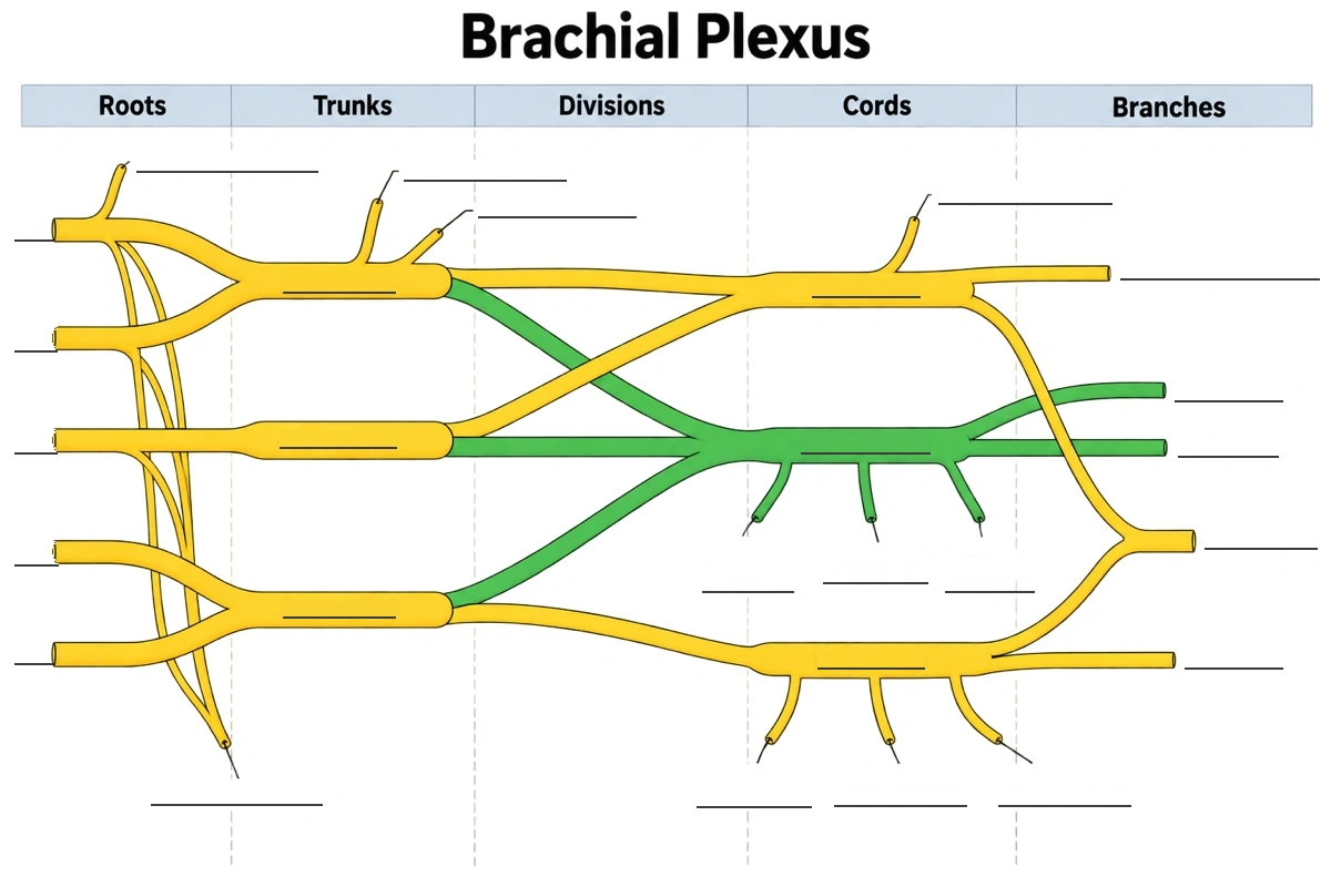
Brachial plexus labeling: the ladder with empty lines
The labeled ladder above with 27 empty answer lines and the five column headers left in. Fill it from the left, then check these four spots:
- 1
Three trunks, five roots
Two roots meet at the top trunk and two at the bottom one; the middle trunk is C7 alone.
- 2
The green cord
The only green cord, fed from all three trunks, is the posterior cord; its two green ends are axillary above radial.
- 3
The V on the right
Where lines from the top and bottom cords meet is the median nerve, musculocutaneous above it, ulnar below.
- 4
Collaterals by level
Off C5 the dorsal scapular, three twigs down to the long thoracic, then suprascapular before nerve to subclavius.
Erb's palsy vs Klumpke's palsy
Erb's palsy is an upper plexus injury of C5 and C6 that weakens the shoulder and elbow; Klumpke's palsy is a lower plexus injury of C8 and T1 that weakens the hand. On a brachial plexus diagram the difference is which end of the ladder is shaded: the upper trunk or the lower trunk.
| Erb's palsy (upper) | Klumpke's palsy (lower) | |
|---|---|---|
| Roots | C5, C6 (sometimes C7) | C8, T1 |
| Part of the plexus | Upper trunk | Lower trunk |
| How it happens | Head and shoulder forced apart: shoulder dystocia at birth, a fall or a motorbike crash onto the shoulder | Arm pulled up above the head: a breech delivery, grabbing a branch while falling |
| Nerves hit hardest | Suprascapular, musculocutaneous, axillary, nerve to subclavius | Ulnar and the medial root of the median |
| Muscles weak | Deltoid, supraspinatus, infraspinatus, biceps, brachialis | Intrinsic muscles of the hand |
| Classic sign | Waiter's tip: arm adducted and medially rotated, elbow extended, forearm pronated | Claw hand: fingers extended at the knuckles and flexed at the finger joints |
| Numb skin | Outer shoulder and lateral arm (C5–C6 dermatomes) | Medial forearm and hand (C8–T1 dermatomes) |
| How common | The commoner of the two | Much rarer; Horner's syndrome if T1 is torn near the spinal cord |
Most birth injuries are upper or upper-and-middle (C5–C7); a pure lower lesion is uncommon, and a total plexus injury takes all five roots. When shading a diagram, mark the roots and the trunk together, then trace which terminal nerves leave the shaded part.
Brachial plexus questions
Where is the brachial plexus located?
At the side of the neck and in the armpit. Its roots come out between the anterior and middle scalene muscles, its trunks cross the base of the posterior triangle of the neck above the first rib, its divisions pass behind the clavicle, and its cords and branches lie in the axilla around the axillary artery before running down the arm.
What are the roots of the brachial plexus?
The anterior rami of the spinal nerves C5, C6, C7, C8 and T1. The posterior rami of the same nerves go to the back muscles and skin, not into the plexus. A small contribution from C4 makes a prefixed plexus, one from T2 a postfixed plexus.
What are the five terminal branches of the brachial plexus?
The musculocutaneous, axillary, radial, median and ulnar nerves. The musculocutaneous comes off the lateral cord, the axillary and radial off the posterior cord, the ulnar off the medial cord, and the median is formed from both the lateral and the medial cords.
What is the mnemonic for the brachial plexus?
Randy Travis Drinks Cold Beer: roots, trunks, divisions, cords, branches. For the terminal nerves in order on the schematic, MARMU (musculocutaneous, axillary, radial, median, ulnar); for the posterior cord, ULTRA (upper subscapular, lower subscapular, thoracodorsal, radial, axillary).
Which cord does the axillary nerve come from?
The posterior cord, with root values C5 and C6. It leaves the axilla through the quadrangular space with the posterior circumflex humeral artery, winds round the surgical neck of the humerus, and supplies the deltoid and teres minor and the skin over the lower deltoid.
Which nerve damage causes a winged scapula?
Usually the long thoracic nerve (C5, C6, C7), which supplies serratus anterior. It comes straight off the roots and runs down the outside of the chest wall, where it is exposed in surgery and to strain; with serratus anterior weak, the medial border of the scapula lifts off the ribs when pushing against a wall.
What does the dorsal scapular nerve supply?
The rhomboid major, rhomboid minor and part of levator scapulae. It is the first branch of the plexus, coming off the C5 root alone and piercing the middle scalene, which is why it sits at the top left of a fully labeled schematic.
What does the thoracodorsal nerve supply?
Latissimus dorsi. It comes off the posterior cord (C6–C8) between the upper and lower subscapular nerves and runs down the posterior wall of the axilla to the muscle; surgeons reuse it as a donor nerve in plexus repair.
Why does an interscalene block miss the ulnar nerve?
Because the injection is made around the C5 to C7 roots and upper trunks, and the local anaesthetic often fails to reach C8 and T1, the roots of the ulnar nerve. It suits shoulder surgery, and it nearly always blocks the phrenic nerve on that side too, which lies on anterior scalene close by.
What is the M in the brachial plexus?
The letter formed by the musculocutaneous, median and ulnar nerves in front of the axillary artery. The lateral cord gives the musculocutaneous nerve and the lateral root of the median, the medial cord gives the medial root and the ulnar nerve, and the two roots meeting make the middle of the M. It is the landmark used to orient a dissection.
Sources
- The Brachial Plexus — TeachMeAnatomy
- Brachial plexus: Anatomy, branches and mnemonics — Kenhub
Keep exploring
Our other diagram tools, and the guides behind them.
Draw your brachial plexus diagram
The full ladder, one cord with its branches, an injury level or the block sites, labeled for your notes or left with answer lines for revision.
Start drawing