
Ear Diagram Maker
Generate labeled ear diagrams: the outer, middle and inner ear in section, the ossicles, the path of sound and unlabeled copies to label. Suited to biology and A&P teaching and study, not for clinical use.
Ear diagram examples
What each prompt drew: the full coronal section of the right ear with the three regions bracketed, an eight-label school ear, the hammer, anvil and stirrup close up against the oval window, and the path of sound in seven numbered steps. Click a card to load its prompt and edit it.
Labeled ear diagram: the parts of the ear and their functions
Each part on the labeled ear diagram, the region it belongs to and what it does, from the outside in.
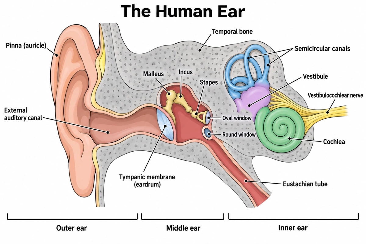
| Part | Region | What it does |
|---|---|---|
| Pinna (auricle) | Outer ear | Elastic-cartilage flap that gathers sound and helps tell whether it comes from in front or behind |
| External auditory canal | Outer ear | Carries sound about 2.5 cm to the eardrum; its glands make earwax |
| Tympanic membrane (eardrum) | Outer–middle border | Thin membrane that vibrates with sound and moves the malleus |
| Malleus (hammer) | Middle ear | First ossicle; its handle is attached to the eardrum |
| Incus (anvil) | Middle ear | Middle ossicle, linking the malleus to the stapes |
| Stapes (stirrup) | Middle ear | Last ossicle; its footplate rocks in the oval window and pushes on the cochlear fluid |
| Oval window | Middle–inner border | Membrane-covered opening where vibration enters the cochlea |
| Round window | Middle–inner border | Bulges out to relieve the pressure wave once it has run through the cochlea |
| Eustachian tube | Middle ear to nasopharynx | Lets air in and out to keep pressure equal on both sides of the eardrum; drains the middle ear |
| Cochlea | Inner ear | Coiled, fluid-filled tube whose hair cells turn vibration into nerve signals for hearing |
| Vestibule | Inner ear | Holds the utricle and saccule, which sense head tilt and straight-line acceleration |
| Semicircular canals | Inner ear | Three fluid-filled loops at right angles that sense head rotation |
| Vestibulocochlear nerve (CN VIII) | Inner ear to brainstem | Carries hearing signals from the cochlea and balance signals from the vestibule and canals |
| Temporal bone | Around all three | The skull bone that houses the ear canal, middle ear and inner ear |
School sheets often merge the three ossicles into one "ear bones" label and call the vestibulocochlear nerve the auditory nerve. Anatomy courses keep them apart and add the vestibule and both windows. Say which set your class uses and only those labels are drawn.
Inner, middle, outer: which ear diagram to draw
Most searches are for one region of the ear, or for one route through it. These are the versions people ask for and the parts each should carry.
Inner ear diagram
The bony labyrinth inside the temporal bone and the membranous labyrinth inside that. Hearing: the cochlea, about two and a half turns, cut across to show the scala vestibuli, the cochlear duct with the organ of Corti on the basilar membrane, and the scala tympani. Balance: the vestibule with the utricle and saccule, and the three semicircular canals at right angles, each with a swollen ampulla. Perilymph fills the bony part, endolymph the membranous part.
Middle ear diagram
The air-filled tympanic cavity between the eardrum and the inner ear: the malleus, incus and stapes, the oval window above the round window on the inner wall, the opening of the Eustachian tube at the front, and the mastoid air cells behind. Fuller versions add the two small muscles — the tensor tympani on the malleus and the stapedius on the stapes, the smallest skeletal muscle in the body.
Parts of the outer ear
The external ear seen from the side, the version many people searching "parts of the outer ear" want: the curved rim or helix, the inner ridge or antihelix, the hollow concha leading into the canal, the small flap in front of it, the tragus, the antitragus opposite, the scaphoid and triangular fossae, and the soft lobule at the bottom, the only part without cartilage.
Ear canal diagram
The external auditory canal, about 2.5 cm long and S-shaped, from the concha to the eardrum. Its outer third has cartilage walls with hairs and the ceruminous glands that make earwax; the inner two thirds run through bone and are lined with thin skin. The eardrum closes it at an angle, so the canal is longer along its floor than along its roof.
Ear bone diagram (the ossicles)
The three ossicles drawn large: the malleus (hammer) with its handle in the eardrum and its head in the attic of the middle ear, the incus (anvil) with its body and long process, and the stapes (stirrup) with its head, two crura and an oval footplate in the oval window. The stapes is about 3 mm long, the smallest bone in the body. Two small synovial joints link the three.
Simple ear diagram
For a first lesson, six to eight labels are enough: pinna, ear canal, eardrum, ear bones, cochlea, semicircular canals, auditory nerve and Eustachian tube. The three bones can share one label, and the vestibule and windows are left out. Keep the cochlea in the inner ear and the tube running down to the throat — the two places a simplified ear most often goes wrong.
Labeled vs unlabeled ear diagram
A labeled ear diagram is the answer key; the unlabeled ear diagram is the same drawing with empty lines, for a quiz or to label from memory. Use one drawing for both so the leader lines match. GCSE and middle-school sheets usually carry 8–10 labels; an A&P lab sheet goes to 14 or more, adding the windows, the vestibule and the nerve.
Ear anatomy diagram with the temporal bone
The anatomy-course version: the ear placed in the petrous part of the temporal bone, with the internal acoustic meatus carrying the vestibulocochlear nerve and the facial nerve beside it, the chorda tympani crossing the middle ear, the mastoid process behind and the Eustachian tube opening into the nasopharynx. Say "left ear" or "right ear" — a coronal section is mirrored between the two.
Path of sound through the ear
The hearing route as numbered steps: the pinna funnels sound into the canal, the eardrum vibrates, the malleus, incus and stapes pass the vibration on, the stapes rocks the oval window, a fluid wave runs through the cochlea and bends hair cells on the basilar membrane, and the cochlear nerve carries the signal to the brainstem and the auditory cortex. High pitches peak near the base of the cochlea, low pitches near its tip.
What are the three parts of the ear?
The ear has three parts: the outer ear (pinna, ear canal and eardrum) collects sound, the middle ear (the malleus, incus and stapes in an air-filled cavity) passes it on, and the inner ear (the cochlea, vestibule and semicircular canals) turns it into nerve signals and senses balance. An ear diagram is usually a coronal section through all three, so the path from the pinna to the vestibulocochlear nerve can be followed in one picture. The figures on this page are drawn for teaching and study, not for clinical use.
Key facts
- The stapes is the smallest bone in the human body, about 3 mm long. With the malleus and incus it carries vibration from the eardrum to the oval window.
- The middle ear raises the pressure of sound about 20 times. The eardrum is roughly 17 times larger than the stapes footplate, and the lever action of the ossicles adds a little more — enough to move fluid instead of air.
- Humans hear about 20 to 20,000 Hz. The basilar membrane sorts pitch by place: high frequencies move it near the base of the cochlea, low frequencies near the tip.
- The cochlea is about two and a half turns, roughly 35 mm long if uncoiled. Its organ of Corti holds about 3,500 inner and 12,000 outer hair cells.
- Half of the inner ear is for balance, not hearing. The utricle and saccule sense tilt and straight-line acceleration; the three semicircular canals sense rotation.
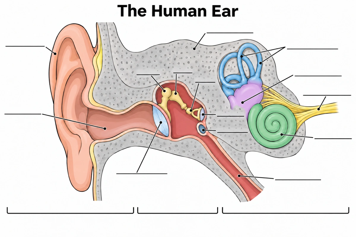
Ear labeling: an unlabeled ear diagram to label
The labeled ear above with fourteen empty answer lines, and the three region brackets left unnamed. The four spots students mix up most:
- 1
Malleus, incus, stapes
Three short lines over the middle ear: hammer at the eardrum, anvil in the middle, stirrup at the window.
- 2
Oval vs round window
Of the two lines on the cochlea's wall, the upper one is the oval window, under the stapes footplate.
- 3
Vestibule vs cochlea
The lilac chamber under the canals is the vestibule; the green coil below and in front of it is the cochlea.
- 4
Bone and brackets
The grey bone around everything is the temporal bone; the brackets read outer, middle and inner ear.
Outer ear vs middle ear vs inner ear
The outer ear carries sound through air, the middle ear carries it through bone, and the inner ear carries it through fluid and turns it into nerve signals. Each region ends at a membrane: the eardrum closes the outer ear, the oval and round windows close the middle ear.
| Outer ear | Middle ear | Inner ear | |
|---|---|---|---|
| Parts | Pinna, ear canal, outer face of the eardrum | Malleus, incus, stapes, tympanic cavity, Eustachian tube | Cochlea, vestibule, semicircular canals |
| Filled with | Air, open to the outside | Air, vented through the Eustachian tube | Fluid: perilymph and endolymph |
| Sound travels as | Pressure waves in air | Vibration of three bones | Fluid waves, then nerve impulses |
| Job | Collect and channel sound | Amplify and pass it on | Detect pitch and loudness; sense balance |
| Hearing loss if it fails | Conductive | Conductive | Sensorineural |
On a diagram, the eardrum is the line between the outer and middle ear and the stapes footplate in the oval window is the line between the middle and inner ear. Brackets that put the eardrum inside the middle ear, or the cochlea beside the ossicles, are the first things to check.
Questions about the ear diagram
What are the parts of the ear?
The outer ear (pinna, ear canal), the eardrum, the middle ear (malleus, incus, stapes and the Eustachian tube) and the inner ear (cochlea, vestibule and semicircular canals), with the vestibulocochlear nerve leading to the brain. A labeled ear diagram for an anatomy course also names the oval and round windows and the temporal bone around them.
What are the parts of the outer ear called?
The pinna (auricle) and the external auditory canal; on the pinna itself, the helix, antihelix, concha, tragus, antitragus, the scaphoid and triangular fossae and the lobule. The eardrum sits at the inner end of the canal and is usually counted as the boundary with the middle ear.
What are the three bones in the ear called?
The malleus (hammer), incus (anvil) and stapes (stirrup), in that order from the eardrum inward. Together they are the auditory ossicles. The malleus is attached to the eardrum, the incus joins it to the stapes, and the stapes footplate fits into the oval window of the cochlea.
Is the eardrum part of the outer or middle ear?
It is the wall between them. Its outer surface faces the ear canal and its inner surface faces the middle-ear cavity, with the malleus handle attached; most diagrams draw the outer-ear bracket ending at the eardrum and the middle-ear bracket starting just behind it.
Which part of the ear controls balance?
The vestibular system of the inner ear: the three semicircular canals, which sense rotation of the head, and the utricle and saccule in the vestibule, which sense tilt and straight-line movement. Their signals travel on the vestibular branch of the vestibulocochlear nerve, alongside the hearing signals from the cochlea.
What does the cochlea do?
It turns sound vibrations into nerve signals. The stapes pushes a wave through the fluid in the coiled cochlea; the wave bends the hair cells of the organ of Corti on the basilar membrane, and they fire the cochlear nerve. Where along the coil the wave peaks tells the brain the pitch: high notes near the base, low notes near the tip.
What does the Eustachian tube do?
It connects the middle ear to the nasopharynx at the back of the nose, so air can enter or leave and the pressure stays equal on both sides of the eardrum. Swallowing or yawning opens it, which is the pop felt on a plane. It also drains the middle ear; in young children it is shorter and more level.
How does sound travel through the ear?
Pinna → ear canal → eardrum → malleus → incus → stapes → oval window → fluid in the cochlea → hair cells → cochlear nerve → brain. It goes as air pressure waves in the outer ear, bone vibration in the middle ear and fluid waves in the inner ear, and leaves as electrical signals.
What should a labeled ear diagram include?
At minimum the pinna, ear canal, eardrum, the three ossicles, the cochlea, the semicircular canals, the Eustachian tube and the auditory nerve. Fuller answer keys add the oval and round windows, the vestibule and the temporal bone, with brackets for the outer, middle and inner ear.
What is the smallest bone in the human body?
The stapes, in the middle ear — about 3 mm long and weighing a few milligrams. Its two arms and oval footplate give it the stirrup shape behind its common name; the footplate is the only part of the ossicle chain that touches the inner ear.
Sources
- 14.1 Sensory Perception (Audition and Equilibrium), in OpenStax Anatomy and Physiology 2e — OpenStax
- How Do We Hear? — National Institute on Deafness and Other Communication Disorders (NIDCD)
Keep exploring
Our other diagram tools, and the guides behind them.
Tools



Draw your ear diagram
The whole ear, one region or the path of sound, labeled or left with empty lines for your class.
Start drawing