
Labeled Digestive System Diagram Maker
Generate labeled digestive system diagrams of the whole tract, a simple ten-label version, the GI tract alone, a flow chart of the path of food and unlabeled copies to label. Each digestive system diagram suits biology class, A&P study or revision notes.
Digestive system diagram examples
Four prompts and the digestive system diagram each one drew: every organ with its job, a ten-label school version, the GI tract alone and a flow chart of the path of food. Pick a card to load its prompt into the box above.
Labeled digestive system diagram with the function of each part
Each label on the digestive system diagram, where the organ sits and its job, in the order food meets it. Food never passes through the accessory organs on the digestive system diagram: the liver, gallbladder, pancreas, tongue and salivary gland.
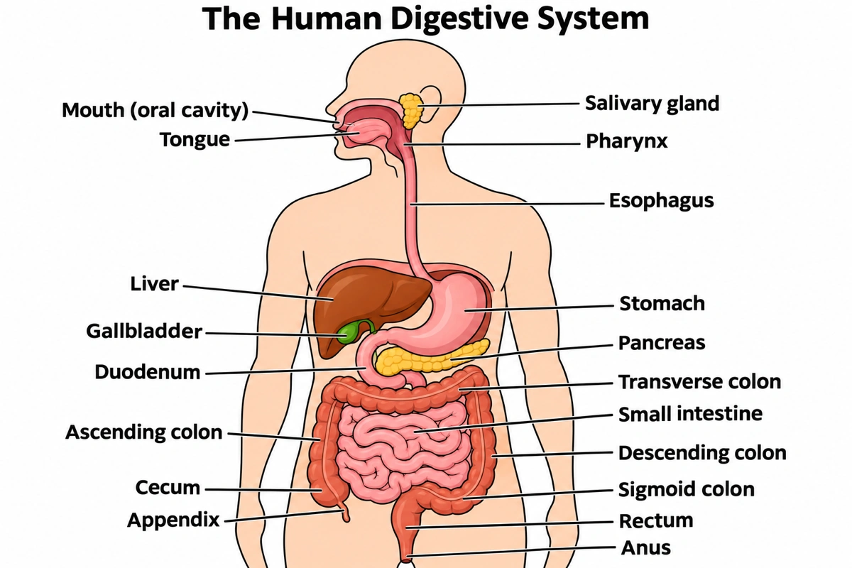
| Part on the digestive system diagram | Where it sits | What it does in digestion |
|---|---|---|
| Mouth (oral cavity) | Head | Teeth grind food and saliva wets it into a bolus; mechanical digestion starts |
| Tongue | Floor of the mouth | Moves food, shapes the bolus and pushes it back to be swallowed |
| Salivary gland | Three pairs around the mouth (parotid, submandibular, sublingual) | Secrete 1 to 1.5 liters of saliva a day; salivary amylase starts starch digestion |
| Pharynx | Behind the mouth and nasal cavity | Shared passage for food and air; swallowing sends the bolus on |
| Esophagus | Down the chest behind the trachea, through the diaphragm | About 25 cm; peristalsis carries the bolus to the stomach |
| Liver | Upper right abdomen, under the diaphragm | Largest gland, about 1.4 kg; makes about 1 liter of bile a day and processes absorbed nutrients |
| Gallbladder | Under the liver | 8 to 10 cm sac that stores and concentrates bile for the duodenum |
| Stomach | Upper left abdomen | Churns food with acid and pepsin into chyme, emptied in 2 to 4 hours |
| Pancreas | Behind the stomach, head in the curve of the duodenum | Over 1 liter of pancreatic juice a day: amylase, lipase, trypsin, nucleases, bicarbonate |
| Duodenum | First 25 cm of the small intestine | Receives chyme, bile and pancreatic juice |
| Small intestine | Coiled mid-abdomen | About 3 m in life; the jejunum and ileum absorb most nutrients through villi |
| Cecum | Lower right abdomen | Pouch where the large intestine begins, past the ileocecal valve |
| Appendix | Hanging from the cecum | About 7.6 cm, rich in lymphoid tissue; food does not pass through it |
| Ascending colon | Right side | Carries residue up while water and salts are absorbed |
| Transverse colon | Across, below the stomach | Absorbs water; gut bacteria ferment what is left |
| Descending colon | Left side | Carries the drying feces down |
| Sigmoid colon | Lower left, S-shaped | Joins the descending colon to the rectum |
| Rectum | Pelvis, the last 20 cm | Stores feces until defecation |
| Anus | End of the tract | Internal (involuntary) and external (voluntary) sphincters pass feces |
A school digestive system diagram has eight to ten labels and calls the colon the large intestine; an A&P answer key names the cecum, appendix, four colon parts and rectum. Name your class's set and the digestive system diagram carries only those labels.
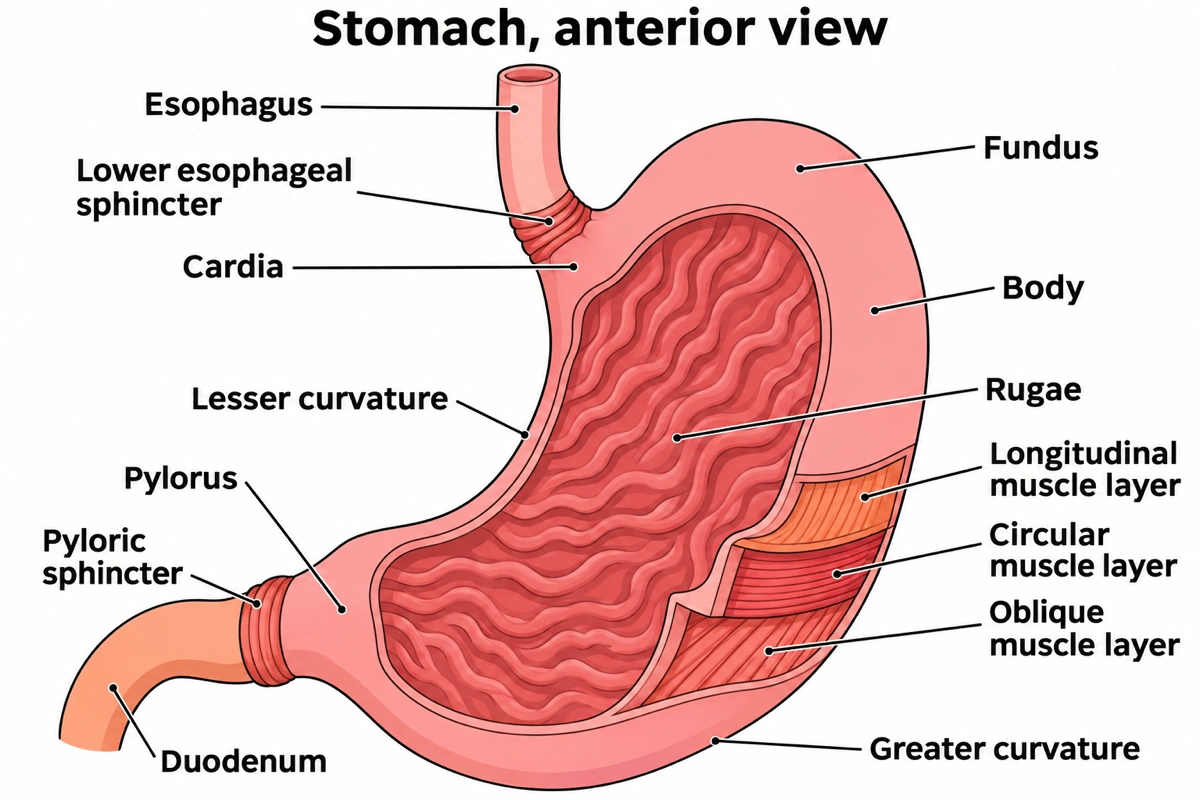
Labeled stomach diagram, the close-up a digestive system diagram leaves out
The J-shaped stomach in front view, its wall opened to show four regions and three muscle layers.
- 1
Cardia and fundus
Food enters the cardia through the lower esophageal sphincter; on a digestive system diagram the fundus is the dome beside it.
- 2
Body and pylorus
The body mixes; the pylorus funnels chyme through the pyloric sphincter to the duodenum.
- 3
Rugae
Folds of the empty lining that flatten as the stomach fills toward 4 liters. A digestive system diagram shows them only in a cutaway.
- 4
Three muscle layers
Longitudinal, circular and an inner oblique layer that lets the stomach churn, labeled outer to inner.
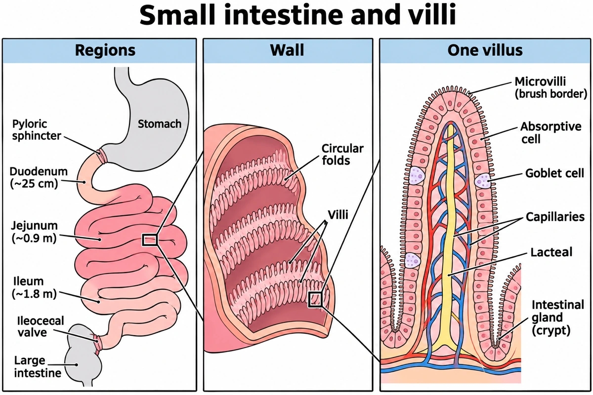
Small intestine diagram, the digestive system down to one villus
Where most food is digested and absorbed, at three scales no whole-body digestive system diagram can show.
- 1
Duodenum, jejunum, ileum
About 25 cm, 0.9 m and 1.8 m in life. On a digestive system diagram the ileum ends at the ileocecal valve.
- 2
Circular folds and villi
Permanent ridges of the lining carpeted with villi 0.5 to 1 mm long.
- 3
Inside a villus
Capillaries take up sugars and amino acids; the central lacteal takes up fats.
- 4
About 200 m² of surface
Folds, villi and microvilli give over 100 times the skin's area, which no digestive system diagram draws to scale.
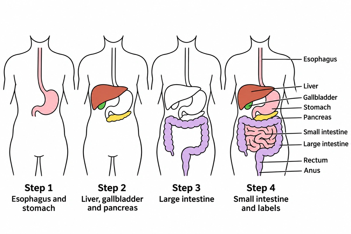
How to draw a digestive system diagram in four steps
Draw the organs of a digestive system diagram in the order they overlap, so nothing has to be erased.
- 1
Esophagus and stomach
Start the digestive system diagram with a tube down the midline and a J-shaped stomach on your right.
- 2
Liver, gallbladder, pancreas
A wedge-shaped liver across the top, larger on your left, the gallbladder under it, the pancreas below the stomach.
- 3
Large intestine
Frame the abdomen with an upside-down U, then run the rectum down to the anus.
- 4
Small intestine and labels
Fill the frame with coils, then label the digestive system diagram with straight lines that do not cross.
Digestive system diagram versions for class, exams and lab practicals
A digestive system diagram can be a ten-label sketch or a single villus. These are the versions searched for most, and what each carries.
Diagram of the human digestive system
The full digestive system diagram: the alimentary canal from mouth to anus plus the salivary glands, liver, gallbladder and pancreas. Textbooks turn the head in profile and draw the trunk from the front, so the liver sits on the viewer's left. The answer key above has nineteen labels; the first digestive system diagram in the gallery writes each organ's job under its name.
Simple digestive system diagram
A simple digestive system diagram has eight to ten labels: mouth, esophagus, stomach, liver, gallbladder, pancreas, small and large intestine, rectum, anus. Shapes are simplified without moving anything: a wedge liver, a J stomach, a frame of colon around the coils.
Gastrointestinal system diagram (GI tract)
A GI tract digestive system diagram shows the tube alone, which is what "GI tract labeled" usually means: mouth, pharynx, esophagus, stomach, duodenum, jejunum, ileum, cecum and appendix, the four parts of the colon, rectum and anus. Leaving the liver and pancreas off keeps the path of food readable.
Digestive system flow chart
Boxes instead of anatomy, mouth to anus, each with one line on what happens there; saliva, bile and pancreatic juice join from side boxes. As a digestive system diagram it answers "how does the digestive system work" in one figure.
Digestive system drawing (easy)
For homework and notes, follow the four-step figure above: esophagus and stomach, liver, the colon frame, then the coils. Keep the liver and appendix on the figure's right side, the two things a quick digestive system diagram sketch most often flips.
Neat labelled diagram of the human digestive system for exams
Exams ask you to "draw a neat labelled diagram of the human digestive system" and mark positions and label count, not art: mouth, salivary glands, esophagus, liver, gallbladder, stomach, pancreas, both intestines, appendix, rectum and anus, with pencil label lines that do not cross. Some papers then ask for the function of two parts of the digestive system diagram, which the table above gives.
Digestive system model labeled
Lab practicals use a plastic torso with removable organs. A matching digestive system diagram is drawn from the front, transverse colon across the abdomen and the liver's right lobe over the gallbladder. Ask for numbered pins instead of names to turn the digestive system diagram into a practice sheet.
Large intestine diagram
Half as long as the small intestine but wider: cecum, appendix, ascending, transverse, descending and sigmoid colon, rectum and anal canal. A detailed digestive system diagram of it adds the teniae coli, three bands of muscle, and the haustra, the pouches they gather the wall into.
Liver, gallbladder and pancreas diagram
The part of a digestive system diagram that shows where bile and enzymes come from: the liver's right and left lobes, hepatic ducts into the common hepatic duct, the cystic duct to the gallbladder, and the common bile duct meeting the pancreatic duct at the hepatopancreatic ampulla in the duodenum.
Cow digestive system diagram
A ruminant's stomach has four compartments: the rumen and reticulum, where microbes digest cellulose and cud is brought up to be rechewed, the omasum, which removes water, and the abomasum, which digests with the cow's own enzymes. A cow digestive system diagram draws them in that order.
What is the digestive system?
The digestive system is the alimentary canal, the tube from the mouth to the anus, together with the accessory organs that serve it: the teeth, tongue, salivary glands, liver, gallbladder and pancreas. It breaks food into molecules small enough to absorb through six processes: ingestion, propulsion, mechanical digestion, chemical digestion, absorption and defecation. A digestive system diagram shows these organs in place so the route of food can be followed in one picture.
Key facts
- The small intestine's absorptive surface is about 200 m², more than 100 times your skin, because folds, villi and microvilli multiply its area over 600-fold.
- About 90 percent of the water entering the gut is absorbed in the small intestine, not the colon, so a digestive system diagram that labels the colon "absorbs water" tells only part of the story.
- Each day the salivary glands make 1 to 1.5 liters of saliva, the liver about 1 liter of bile and the pancreas over 1 liter of pancreatic juice.
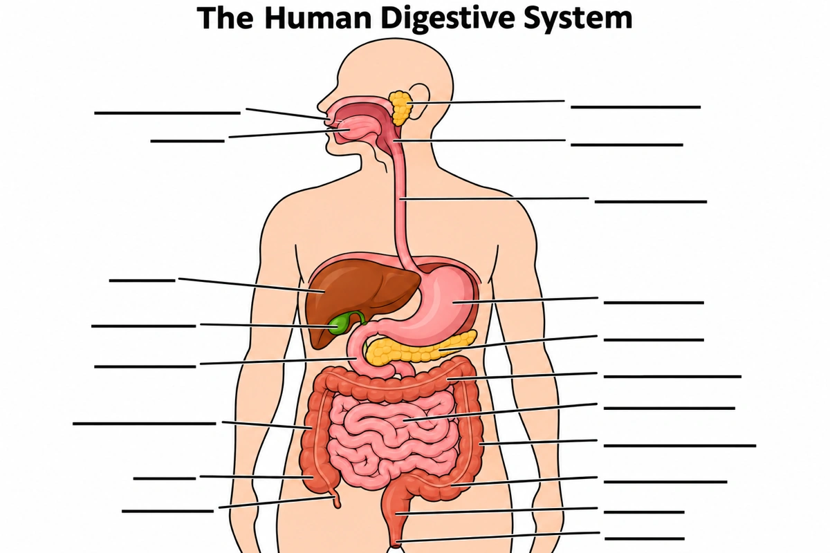
Blank digestive system diagram to label
The digestive system diagram answer key above with all nineteen labels replaced by empty lines. Where students lose marks:
- 1
Which side is which
On a front-view digestive system diagram the liver and gallbladder are on your left, the stomach on your right.
- 2
Duodenum or pancreas
On the digestive system diagram, the C-shaped tube leaving the stomach is the duodenum; the gland in its curve is the pancreas.
- 3
Cecum and appendix
At the bottom left of the digestive system diagram, the pouch is the cecum and the thin tube hanging from it the appendix.
- 4
Four parts of the colon
Up is ascending, across transverse, down descending, and the S-bend into the rectum sigmoid, on any front-view digestive system diagram.
Alimentary canal vs accessory organs of the digestive system
Food passes through the alimentary canal but never through the accessory organs, which send their secretions in through ducts. That is why a gastrointestinal system diagram can leave out the liver and pancreas while a full digestive system diagram cannot.
| Alimentary canal (GI tract) | Accessory digestive organs | |
|---|---|---|
| Organs | Mouth, pharynx, esophagus, stomach, small and large intestine, rectum, anus | Teeth, tongue, salivary glands, liver, gallbladder, pancreas |
| Does food pass through? | Yes, mouth to anus | No, except the teeth and tongue, which work inside the mouth |
| Main job | Moves, digests and absorbs food; expels waste | Supplies saliva, bile and pancreatic juice |
| Wall | Mucosa, submucosa, muscularis, serosa | Glandular tissue drained by ducts |
| On a digestive system diagram | One continuous tube, labeled region by region | Beside the tube, joined by ducts |
Color the canal in one family of tints and the accessory organs in another, and the two groups read at a glance. The first digestive system diagram in the gallery uses pinks and purples for the tract and brown, green and yellow for the liver, gallbladder and pancreas.
Questions about the digestive system diagram
What are the organs of the digestive system?
The alimentary canal (mouth, pharynx, esophagus, stomach, small intestine, large intestine, rectum, anus) and the accessory organs (teeth, tongue, salivary glands, liver, gallbladder, pancreas). A school digestive system diagram usually labels ten of them.
In what order does food pass through the digestive system?
Mouth → pharynx → esophagus → stomach → duodenum → jejunum → ileum → cecum → ascending, transverse, descending and sigmoid colon → rectum → anus. Bile and pancreatic juice join in the duodenum, so a digestive system diagram draws the liver and pancreas beside the route, not on it.
Why is the liver on the left side of a digestive system diagram?
Because the figure faces you: the liver is on the body's right, which is the viewer's left. The same rule puts the stomach on the right and the cecum and appendix low on the left of a front-view digestive system diagram.
What is the difference between the digestive system and the GI tract?
The GI tract is the tube food travels through; the digestive system is that tube plus the accessory organs. A gastrointestinal system diagram can drop the liver, gallbladder and pancreas, which a digestive system diagram includes.
Where does most digestion and absorption happen?
In the small intestine. Bile and pancreatic enzymes finish digestion there, and most carbohydrates and proteins are absorbed in the jejunum, so it is the organ to annotate most on a digestive system diagram.
What does the pancreas do in digestion?
It sends over a liter of pancreatic juice a day into the duodenum: amylase, lipase, trypsin, chymotrypsin, nucleases and bicarbonate. On a digestive system diagram it lies behind the stomach, its head in the curve of the duodenum.
How long does food take to pass through the digestive system?
Roughly 17 to 33 hours, adding up the textbook figures. The stomach empties in 2 to 4 hours, the small intestine in 3 to 5, and residue stays 12 to 24 hours in the large intestine; a digestive system diagram drawn as a flow chart can carry these times.
What are the four regions of the stomach?
The cardia, fundus, body and pylorus. The pylorus ends at the pyloric sphincter, which lets chyme into the duodenum; the stomach close-up above labels all four, while a whole-body digestive system diagram labels only "stomach".
How many labels should a digestive system diagram have?
About eight to ten for middle school or GCSE, fifteen to twenty for anatomy and physiology. The digestive system diagram maker draws exactly the labels your prompt lists.
How do you draw a digestive system diagram easily?
Draw the organs in the order they overlap: esophagus and stomach, liver, the colon frame, then the small intestine coils. The four-step figure above follows that order, and a generated digestive system diagram can serve as your tracing reference.
Is the appendix part of the digestive system?
Yes, a small tube on the cecum, though food does not pass through it. A full digestive system diagram labels it at the lower right of the abdomen, on the viewer's left.
What does a cow digestive system diagram show?
A cow digestive system diagram shows one stomach with four compartments in sequence: rumen, reticulum, omasum and abomasum. Microbes in the first two break down cellulose, the omasum removes water and the abomasum digests with the cow's own enzymes.
Sources
- 23.1 Overview of the Digestive System, in OpenStax Anatomy and Physiology 2e — OpenStax
- 23.3 The Mouth, Pharynx, and Esophagus, in OpenStax Anatomy and Physiology 2e — OpenStax
- 23.4 The Stomach, in OpenStax Anatomy and Physiology 2e — OpenStax
- 23.5 The Small and Large Intestines, in OpenStax Anatomy and Physiology 2e — OpenStax
- 23.6 Accessory Organs in Digestion, in OpenStax Anatomy and Physiology 2e — OpenStax
- 23.7 Chemical Digestion and Absorption, in OpenStax Anatomy and Physiology 2e — OpenStax
- 34.1 Digestive Systems, in OpenStax Biology 2e — OpenStax
Keep exploring
Our other diagram tools, and the guides behind them.
Tools

Make your digestive system diagram
A digestive system diagram of the whole tract, the GI tube, one organ or a flow chart, labeled or blank for your class.
Start drawing