
Labeled Brain Diagram Maker for Lobes and Parts of the Brain
Generate a brain diagram labeled with the lobes, sulci, cerebellum, pons and medulla, a midsagittal section, the limbic system or the cortex's functional areas, plus a blank copy to label. Made for biology, psychology and A&P classes.
Brain diagram examples
Four brain diagram renders and their prompts: the four lobes with their functions, the pons, medulla and cerebellum, the limbic system in medial view and the functional areas of the cortex. Pick one to copy its prompt into the box, then rename or drop labels.
Labeled brain diagram with the parts of the brain and their functions
Each part named on the labeled brain diagram, where to find it and what it does, from the front of the cerebrum down to the spinal cord.
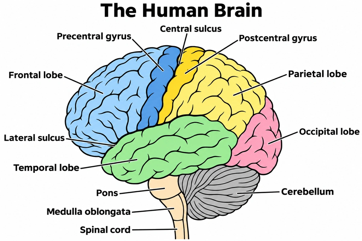
| Part | Where it is on the brain diagram | What it does |
|---|---|---|
| Frontal lobe | Front of the cerebrum, ahead of the central sulcus | Planning, judgment, personality and voluntary movement; Broca's area for speech production sits in its lower part, usually on the left |
| Parietal lobe | Top and upper side, behind the central sulcus | Touch, temperature, pain and the sense of where the body is in space |
| Temporal lobe | Lower side of the cerebrum, below the lateral sulcus | Hearing, language comprehension (Wernicke's area) and memory; the hippocampus and amygdala lie on its inner side |
| Occipital lobe | Back of the cerebrum, above the cerebellum | Vision: the primary visual cortex takes in signals from the eyes |
| Central sulcus | Groove running down from the top, between the frontal and parietal lobes | Landmark on any brain diagram that separates the motor strip in front from the touch strip behind |
| Lateral sulcus | Deep groove running back above the temporal lobe | Separates the temporal lobe from the frontal and parietal lobes; the insula lies hidden in its depth |
| Precentral gyrus | Ridge directly in front of the central sulcus | Primary motor cortex: its upper motor neurons start voluntary movement of skeletal muscle |
| Postcentral gyrus | Ridge directly behind the central sulcus | Primary somatosensory cortex (Brodmann areas 1, 2 and 3): touch, pressure, pain, itch and body position |
| Cerebellum | Under the occipital lobe, behind the pons and medulla | Compares motor commands with sensory feedback to keep balance and movement coordinated |
| Pons | Rounded bulge in front of the cerebellum | Main connection between the cerebrum and the cerebellum; with the medulla it regulates breathing and heart rate |
| Medulla oblongata | Below the pons, just above the spinal cord | Automatic control of breathing, heart rate and blood pressure |
| Spinal cord | Bottom of the brain diagram, below the medulla | Carries sensory signals up from the body and motor commands down to it |
On a lateral brain diagram the midbrain is hidden behind the temporal lobe, so only the pons and medulla show. The sagittal brain diagram below adds it, with the thalamus, hypothalamus and corpus callosum.
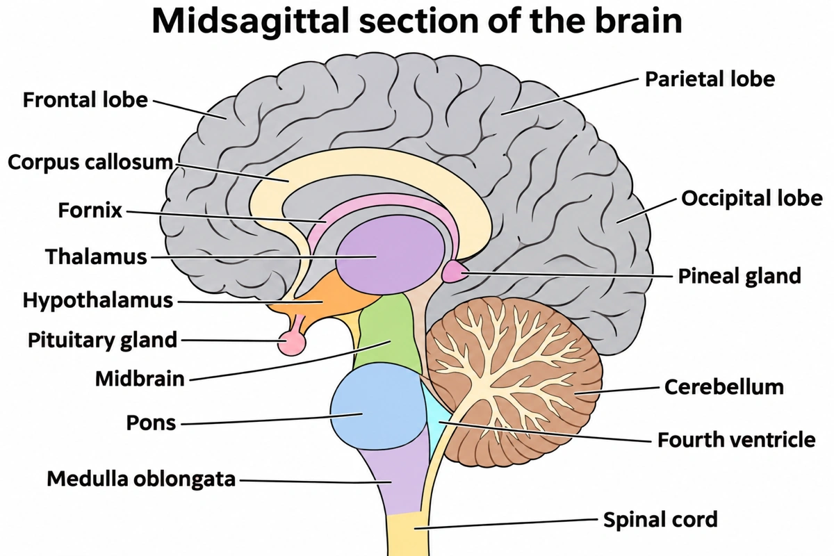
Sagittal section of the brain, labeled
Cut down the midline, the brain diagram shows what the side view hides.
- 1
Corpus callosum and fornix
On a sagittal brain diagram, the pale arch joining the two hemispheres, with the thin fornix under it.
- 2
Thalamus and hypothalamus
The relay for every sense but smell, and below it the hypothalamus holding the pituitary.
- 3
Down to the spinal cord
Midbrain, pons and medulla oblongata in one column.
- 4
Cerebellum
The branching white arbor vitae, behind the fourth ventricle.
Which brain diagram each lesson asks for
Most people want a brain diagram of one view or of one system inside it. These are the versions they look for, the parts each brain diagram should carry and what to put in the prompt to get it.
Lobes of the brain diagram
A side-view brain diagram with the frontal, parietal, temporal and occipital lobes in four colours. The central sulcus divides frontal from parietal and the lateral sulcus sets the temporal lobe apart; the occipital lobe has no sharp border on the side view, so a brain diagram takes its edge from the parieto-occipital sulcus on the inner surface. Some atlases add the insula, folded inside the lateral sulcus, as a fifth lobe.
Cerebrum diagram
A brain diagram of the cerebrum alone: two cerebral hemispheres split by the longitudinal fissure and joined by the corpus callosum, a thin outer cortex of gray matter over white matter. A coronal brain diagram adds the basal nuclei: the caudate beside the lateral ventricle, the putamen below it across the internal capsule, and the globus pallidus just medial to the putamen.
Sagittal section of the brain
The inner brain labeled diagram most courses test, a midline cut showing the corpus callosum, fornix, thalamus, hypothalamus with the pituitary, pineal gland, midbrain, pons, medulla and the cerebellum's arbor vitae. Ask for a "midsagittal" brain diagram to get the exact midline.
Brain stem diagram
This brain diagram covers the midbrain, pons and medulla oblongata from the thalamus down to the spinal cord, with the cerebellum attached behind. An anterior view shows cranial nerves III to XII leaving it; a posterior view, with the cerebellum removed, shows the four colliculi on the midbrain roof and the floor of the fourth ventricle.
Limbic system diagram
A medial-view brain diagram with the hippocampus (forming new long-term memories), the amygdala (emotion, especially fear), the hypothalamus, the cingulate gyrus, the fornix arching from the hippocampus to the mammillary body, and the thalamus. Smell reaches the limbic system directly, which is why the olfactory bulb belongs on the brain diagram.
Cerebral cortex diagram with functional areas
A functional brain diagram maps the cortex by job rather than by lobe: prefrontal cortex, premotor cortex, the primary motor cortex on the precentral gyrus, the primary somatosensory cortex on the postcentral gyrus, Broca's area, Wernicke's area, the primary auditory cortex (Brodmann 41 and 42) and the primary visual cortex (Brodmann 17 and 18).
Simple brain diagram
For a first lesson or a quick brain drawing, a simple brain diagram needs only five to seven labels: cerebrum, the four lobes, cerebellum and brainstem. Leave the sulci and gyri off the brain diagram, keep the cerebellum tucked under the back of the cerebrum and the stem running down in front of it.
Brain diagram for psychology
An intro psychology brain diagram sorts the parts into forebrain, midbrain and hindbrain: the cortex lobes, thalamus, hypothalamus, pituitary and limbic system in front; the reticular formation, substantia nigra and ventral tegmental area in the middle; the medulla, pons and cerebellum behind. Add the corpus callosum and Broca's and Wernicke's areas to the brain diagram for split-brain and language questions.
Sheep brain diagram
For the dissection lab. A sheep brain diagram has the same parts as a human one, but the cerebrum is smaller and the organ lies front to back rather than top to bottom. The ventral brain diagram shows two prominent olfactory bulbs at the front, the X of the optic chiasma with the pituitary beneath it; the cut half shows the corpus callosum, lateral and third ventricles, fornix, thalamus and arbor vitae.
Inferior view of the brain
A brain diagram from below: the olfactory bulbs and tracts under the frontal lobes, the optic chiasm, the pituitary stalk and mammillary bodies, the temporal lobes on either side, then the pons and medulla with the cranial nerves leaving them, and the cerebellar hemispheres at the back.
Brain ventricles diagram
A ventricles brain diagram shows the four fluid-filled cavities: two C-shaped lateral ventricles deep in the cerebrum, joined by the interventricular foramina to the third ventricle between the two halves of the diencephalon, the cerebral aqueduct through the midbrain, and the fourth ventricle between the pons and the cerebellum. Choroid plexus in all four makes the cerebrospinal fluid.
What are the main parts of the brain?
The brain has three main parts: the cerebrum, the large folded upper part split into left and right hemispheres and four lobes; the cerebellum, tucked under the back of the cerebrum; and the brainstem (midbrain, pons and medulla oblongata), which runs down into the spinal cord. Deep between the two hemispheres sits the diencephalon, the thalamus and hypothalamus. A brain diagram usually shows the outside from the side and the inside as a midsagittal section.
Key facts
- Laid flat, the cerebral cortex would cover about one square meter. Folding it into gyri (ridges) and sulci (grooves) is how that much gray matter fits inside the skull.
- The cerebellum is about 10 percent of the brain's mass.
- Every sense except smell passes through the thalamus before it reaches the cerebral cortex; smell goes straight to the cerebrum.
- Brodmann divided the cortex into 52 areas by its microscopic structure; areas 17 and 18 handle vision, 41 and 42 hearing, and 1, 2 and 3 touch.
- The corpus callosum carries about 200 million axons between the two hemispheres. Each hemisphere controls the opposite side of the body.
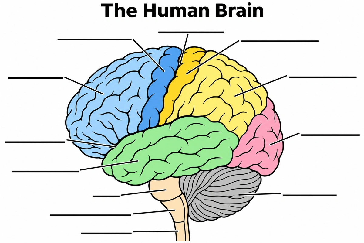
Brain anatomy labeling with an unlabeled brain diagram
The labeled brain diagram above with its twelve labels replaced by empty answer lines. The four pairs students mix up most:
- 1
Central vs lateral sulcus
On the blank brain diagram, the groove running down from the top is central; the one running back above the temporal lobe is lateral.
- 2
Precentral vs postcentral gyrus
Pre means in front: on this brain diagram the motor strip is ahead of the central sulcus, the touch strip behind it.
- 3
Parietal vs occipital lobe
The occipital lobe is only the back tip above the cerebellum; everything between it and the central sulcus on the brain diagram is parietal.
- 4
Pons vs medulla
On the brain diagram the pons is the rounded bulge; the narrower medulla below it joins the spinal cord.
Cerebrum vs cerebellum vs brainstem
The cerebrum thinks, senses and starts movement, the cerebellum coordinates movement, and the brainstem keeps the body alive and connects the cerebrum to the spinal cord. On any brain diagram the cerebrum is the large folded mass on top, the cerebellum the finely ridged one beneath its back, and the narrow stalk in front of the cerebellum is the brainstem.
| Cerebrum | Cerebellum | Brainstem | |
|---|---|---|---|
| Position on a brain diagram | Top and front, the bulk of the organ | Under the occipital lobe, behind the pons | Below the cerebrum, in front of the cerebellum |
| Parts | Two hemispheres, four lobes, cortex, basal nuclei | Two cerebellar hemispheres, vermis, arbor vitae | Midbrain, pons, medulla oblongata |
| Surface | Large gyri and sulci | Fine parallel folds | Smooth white matter outside |
| Main job | Thought, memory, language, senses, voluntary movement | Balance, posture, coordination, motor learning | Breathing, heart rate, blood pressure, sleep and wakefulness, cranial nerves |
| If damaged | Depends on the lobe: weakness, numbness, aphasia, vision loss | Clumsy, unsteady movement (ataxia) | Can stop breathing or consciousness |
Brain diagrams often get the cerebellum's place wrong: it sits under the occipital lobe and behind the pons, never in front of it. The pons should bulge forward on the front of the stem, the medulla taper below it into the spinal cord.
Questions about the brain diagram
What are the parts of the brain on a labeled brain diagram?
A labeled brain diagram in side view shows the cerebrum with its frontal, parietal, temporal and occipital lobes, the central and lateral sulci, the cerebellum, the pons and the medulla oblongata above the spinal cord. A sagittal brain diagram adds the corpus callosum, thalamus, hypothalamus, pituitary gland and midbrain.
What are the 4 lobes of the brain and their functions?
Frontal (planning, judgment, voluntary movement and speech production), parietal (touch, temperature, pain and body position), temporal (hearing, language comprehension and memory) and occipital (vision). Each hemisphere has all four, so a left-side brain diagram and a right-side one carry the same lobe labels.
What is the largest part of the brain?
The cerebrum. It makes up most of the mass and covers the diencephalon and most of the brainstem, which is why a lateral brain diagram is mostly cerebrum, with the cerebellum, pons and medulla showing only underneath.
What does the brainstem do?
It links the cerebrum and cerebellum to the spinal cord and runs the automatic functions: the medulla and pons regulate breathing, heart rate and blood pressure, and the reticular formation running through it controls sleep and wakefulness. On a brain diagram it is the stalk under the cerebrum, and every pathway between the cerebrum and the spinal cord passes through it.
What does a midsagittal brain diagram show?
The inside of one hemisphere along the midline: the corpus callosum, fornix, thalamus, hypothalamus, pituitary and pineal glands, the midbrain, pons and medulla, the fourth ventricle and the cerebellum's arbor vitae. It is the brain diagram to use for any structure a side view hides.
Which part of the brain controls emotions?
The limbic system, chiefly the amygdala, with the hippocampus and the hypothalamus; a limbic system brain diagram shows all three in medial view. The amygdala ties emotional meaning to experiences and memories, the hippocampus forms new long-term memories, and the hypothalamus drives the body's response through the autonomic nervous system and hormones.
Which part of the brain controls speech?
Broca's area in the lower frontal lobe produces speech, and Wernicke's area in the upper temporal lobe understands it; in most people both are on the left side. A brain diagram therefore shows both on the left hemisphere. Damage to Broca's area makes speaking hard; damage to Wernicke's area leaves fluent speech that makes little sense.
Does the left side of the brain control the right side of the body?
Yes. Each hemisphere controls movement and sensation on the opposite side of the body, because most motor and sensory pathways cross over in the medulla or spinal cord. The two hemispheres share information through the corpus callosum, the thick arch at the centre of a sagittal brain diagram.
Is the insula a fifth lobe of the brain?
Some atlases count it as one. The insula is a fold of cortex hidden deep inside the lateral sulcus, covered by the frontal, parietal and temporal lobes, so it never shows on an ordinary side view; a brain diagram reveals it only by pulling the temporal lobe down or in a coronal section.
How many labels should a brain diagram for school have?
Six to eight for a middle-school brain diagram (cerebrum, four lobes, cerebellum, brainstem), twelve to fifteen for high-school biology or A&P. The larger set adds the sulci, gyri, pons, medulla, corpus callosum, thalamus and hypothalamus.
Keep exploring
Our other diagram tools, and the guides behind them.
Make a brain diagram for your class
A brain diagram of the lobes, a sagittal section or the limbic system, labeled as your course names them or left blank to label.
Start drawing