
RAAS Diagram
Describe the renin–angiotensin–aldosterone system you need and get a clean pathway diagram — the trigger at the top, renin, angiotensin I and II and ACE in order, the effects of angiotensin II fanning out to a rise in blood pressure, the negative feedback and the drug targets.
RAAS diagram examples
Real renders of the prompts shown: the full RAAS with the five actions of angiotensin II and its feedback loop, a seven-step overview, the drug targets and a blank version. Click one to load and edit its prompt.
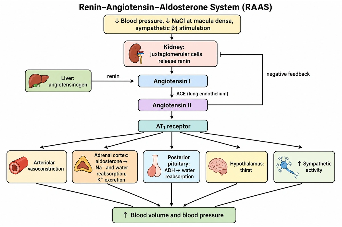
From a fall in pressure to a rise in volume
Read top to bottom; each step is one box in the figure.
- 1
Trigger → renin
Low pressure or low NaCl makes the kidney release renin.
- 2
Angiotensin I → II
Renin and lung ACE turn liver angiotensinogen into angiotensin II.
- 3
Five effects via AT1
Vasoconstriction, aldosterone, ADH, thirst, sympathetic activity.
- 4
Feedback
Angiotensin II inhibits renin, the bar that closes the loop.
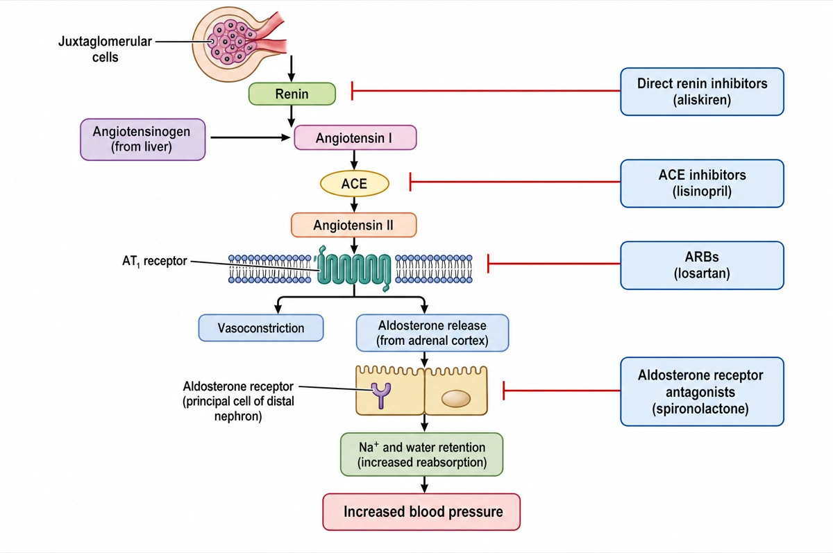
Where the four drug classes cut the chain
Each blocker is a bar on the step it stops.
- 1
ACE inhibitors
Bar on the ACE arrow: less angiotensin II is made.
- 2
ARBs
Bar between angiotensin II and its AT1 effects.
- 3
Renin and MR blockers
Aliskiren stops the first step; spironolactone the aldosterone arm.
What is the renin–angiotensin–aldosterone system?
The renin–angiotensin–aldosterone system (RAAS) is the hormone cascade that raises blood pressure and blood volume: when pressure falls, kidney juxtaglomerular cells release renin, which turns angiotensinogen from the liver into angiotensin I; angiotensin-converting enzyme (ACE), mainly in the lungs, turns that into angiotensin II, which constricts arterioles and releases aldosterone and ADH. Aldosterone makes the kidney keep Na⁺ and, with it, water.
Key facts
- Three signals release renin. Low pressure in the afferent arteriole, less NaCl reaching the macula densa, and sympathetic nerve stimulation through β-receptors all make juxtaglomerular cells secrete renin; circulating angiotensin II feeds back to suppress it.
- Renin and ACE are the two cutting steps. Renin cleaves angiotensinogen into the 10-amino-acid angiotensin I; ACE removes two more amino acids to give the active 8-amino-acid angiotensin II.
- Angiotensin II acts mainly through the AT1 receptor. Vasoconstriction, Na⁺ reabsorption and aldosterone secretion all run through AT1 — the receptor that ARBs block. Angiotensin II also triggers thirst, ADH release and sympathetic discharge.
- The fast arm and the slow arm. Angiotensin II raises pressure at once by vasoconstriction; aldosterone raises it more slowly by making the distal tubule and collecting duct reabsorb Na⁺ (water follows) and excrete K⁺.
- Four drug classes block the chain. Direct renin inhibitors (aliskiren) act on renin, ACE inhibitors (lisinopril) on ACE, angiotensin II receptor blockers (losartan) on the AT1 receptor and aldosterone receptor antagonists (spironolactone) on the aldosterone receptor; an ACE inhibitor and an ARB should not be combined.
How does the RAAS diagram maker work?
Describe the diagram
Say what you need — the full cascade, a simple overview, the drug targets, the negative feedback, or a blank worksheet. Name the steps and organs you want and only those are drawn.
Pick a style
The default is a clean flat textbook plate: the trigger at the top, one box per hormone or enzyme, each organ named beside its step, arrows down to the rise in blood pressure and inhibition bars for feedback and drugs. Or apply watercolor, ink line art and other finishes.
Refine and export
The result opens in your workspace, where you can redraw regions, edit labels on the canvas, upscale and export in high resolution.
Components of the renin–angiotensin–aldosterone system
The steps a labeled RAAS diagram is normally expected to show, where each one is made, what it does and what comes next. This is the default content unless you ask for something else.
| Component | Made in or released from | What it does | Next step |
|---|---|---|---|
| Macula densa | Distal tubule, at the juxtaglomerular apparatus | Senses less NaCl in the tubular fluid | Signals the juxtaglomerular cells |
| Renin | Juxtaglomerular cells of the afferent arteriole | Enzyme that cleaves angiotensinogen | Angiotensin I |
| Angiotensinogen | Liver | Inactive precursor circulating in plasma | Cut by renin |
| Angiotensin I | Blood plasma | Inactive 10-amino-acid peptide | Converted by ACE |
| ACE (angiotensin-converting enzyme) | Endothelium, mainly in the lungs | Converts angiotensin I into angiotensin II | Angiotensin II |
| Angiotensin II | Blood plasma | Constricts arterioles; releases aldosterone and ADH; triggers thirst and sympathetic discharge | AT1 receptors on vessels, adrenal cortex and brain |
| Aldosterone | Adrenal cortex | Na⁺ reabsorption (water follows) and K⁺ excretion in the distal tubule and collecting duct | Blood volume rises |
| ADH (vasopressin) | Posterior pituitary | Water reabsorption in the collecting duct; constricts peripheral vessels | Blood volume rises |
| Natriuretic peptides (ANP, BNP) | Heart atria and ventricles | Counter the RAAS: suppress renin, aldosterone and ADH | Blood pressure falls |
RAS and RAAS name the same system: renin–angiotensin system (RAS) stresses the renin → angiotensin steps, RAAS names aldosterone as well. Name the steps you need and only those are drawn.
ACE inhibitors vs ARBs: where does each act in the RAAS?
ACE inhibitors block the enzyme that turns angiotensin I into angiotensin II; angiotensin II receptor blockers (ARBs) let angiotensin II form but stop it acting on its AT1 receptor. Both lower blood pressure and both are first-line choices for hypertension, but they are not given together.
| ACE inhibitor | ARB | |
|---|---|---|
| Examples | Lisinopril, ramipril | Losartan |
| Step blocked | Angiotensin I → angiotensin II (ACE) | Angiotensin II → AT1 receptor |
| Where to draw the bar | On the ACE arrow | Between angiotensin II and its effects |
| Effect on the cascade | Less angiotensin II is made | Angiotensin II is made but cannot act through AT1 |
| Why one is swapped for the other | Cough or angioedema can make it intolerable | Used when an ACE inhibitor causes cough or angioedema |
| In pregnancy | Contraindicated | Contraindicated |
| Together | Not combined with an ARB | Not combined with an ACE inhibitor |
Two more drug classes act on the same chain: direct renin inhibitors such as aliskiren block renin at the top, and aldosterone receptor antagonists such as spironolactone block aldosterone at the bottom.
Where are RAAS diagrams used?
RAAS diagrams are used mostly in physiology, nursing and pharmacology teaching — blood pressure control, kidney function, antihypertensive drugs — and as figures in cardiovascular and renal papers. The level of detail follows the reader: six boxes for a first lesson, every organ, receptor and drug target for a pharmacology exam.
Physiology and nursing courses
Blood pressure regulation and kidney function units, where the RAAS is drawn from the trigger to aldosterone, often as a blank diagram to complete.
Pharmacology
Showing where the antihypertensive classes act: direct renin inhibitors, ACE inhibitors, ARBs and aldosterone receptor antagonists, each as an inhibition bar on its step.
Clinical teaching
Explaining how the body answers blood loss, dehydration or heart failure, why blocking it lowers blood pressure, and why spironolactone spares potassium.
Research figures and posters
Cardiovascular, renal and endocrine papers, reviews and posters, from the classical cascade to the ACE2 and angiotensin-(1–7) branch.
Common questions about the RAAS
How do I draw a RAAS diagram?
Start at the top with the trigger (low blood pressure), then draw the kidney releasing renin, renin turning angiotensinogen from the liver into angiotensin I, ACE in the lungs making angiotensin II, and angiotensin II fanning out to vasoconstriction, aldosterone, ADH and thirst, all converging on a rise in blood pressure. With this maker you describe the version you need and it is drawn top to bottom in that order.
What are the steps of the renin–angiotensin–aldosterone system?
Low pressure → juxtaglomerular cells release renin → renin converts angiotensinogen to angiotensin I → ACE converts angiotensin I to angiotensin II → angiotensin II constricts arterioles and releases aldosterone and ADH → the kidney keeps Na⁺ and water → blood volume and pressure rise. Rising angiotensin II then suppresses renin.
What triggers renin release?
A fall in pressure sensed in the afferent arteriole, less NaCl reaching the macula densa, and sympathetic stimulation through β-receptors. Angiotensin II itself inhibits renin release, closing the loop.
Where is ACE found?
Mainly on the endothelial cells of the lungs, which is where most angiotensin I is converted, but also in the kidneys and the brain. Most diagrams draw the lungs beside the ACE step.
What does angiotensin II do?
It constricts arterioles, stimulates the adrenal cortex to release aldosterone, releases ADH from the posterior pituitary, triggers thirst and increases sympathetic discharge. Together these raise blood volume and blood pressure.
What does aldosterone do in the RAAS?
Aldosterone makes the distal tubule and collecting duct reabsorb Na⁺ and excrete K⁺; water follows the sodium, so blood volume and pressure rise. It is the slow arm of the system, where angiotensin II vasoconstriction is the fast one.
Where do ACE inhibitors and ARBs act?
ACE inhibitors (lisinopril, ramipril) block the conversion of angiotensin I to angiotensin II; ARBs (losartan) block angiotensin II at its AT1 receptor. Direct renin inhibitors (aliskiren) act one step higher and aldosterone receptor antagonists (spironolactone) at the end of the chain.
How is the RAAS switched off?
By negative feedback: angiotensin II inhibits renin release, and as pressure and NaCl delivery recover the triggers disappear. Natriuretic peptides from the stretched heart also suppress renin, aldosterone and ADH.
Is renin a hormone or an enzyme?
Renin is an enzyme — a protease that cleaves angiotensinogen — although some sources call it a hormone because it is secreted into the blood and drives a hormone system. Diagrams usually draw it as the enzyme beside the angiotensinogen → angiotensin I arrow.
How is this different from a generic AI image tool or a blank diagram editor?
The pathway rules are built in. Every render is instructed to read top to bottom from the trigger to the effect, draw each hormone and enzyme as its own node in its true order, show activation as arrows and inhibition as flat-headed bars, and spell every name exactly. Check the result against your course before printing; the canvas lets you move, relabel or redraw any part.
Can I use the diagrams for teaching or publication?
Yes — diagrams you generate are yours to use in lecture slides, handouts, exam papers, posters and papers. Export high-resolution raster up to 4K depending on the model.
Sources
- Homeostatic Regulation of the Vascular System — Anatomy and Physiology 2e, section 20.4 — OpenStax
- Endocrine Regulation of Kidney Function — Anatomy and Physiology 2e, section 25.8 — OpenStax
- Hypertension — MSD Manual Professional Edition
- Metabolism of Angiotensinogen to Angiotensins (R-HSA-2022377) — Reactome
Keep exploring
Our other diagram tools, and the guides behind them.

Make your RAAS diagram
From one line of description to a labeled, printable renin–angiotensin–aldosterone diagram in about a minute.
Start drawing