
Cell Cycle Diagram
Describe the cell cycle you need and get a clean circular diagram — G1, S, G2 and M in order with arcs sized to the time each phase takes, G0 to one side, the three checkpoints and the cyclin–CDK pairs that drive each step. Or view the labeled and blank versions below, free.
Cell cycle diagram examples
Real renders of the prompts shown: a labeled cell cycle with phases timed to 24 hours, a simple worksheet, the checkpoints with their cyclin–CDK complexes and a blank version. Click one to load and edit its prompt.
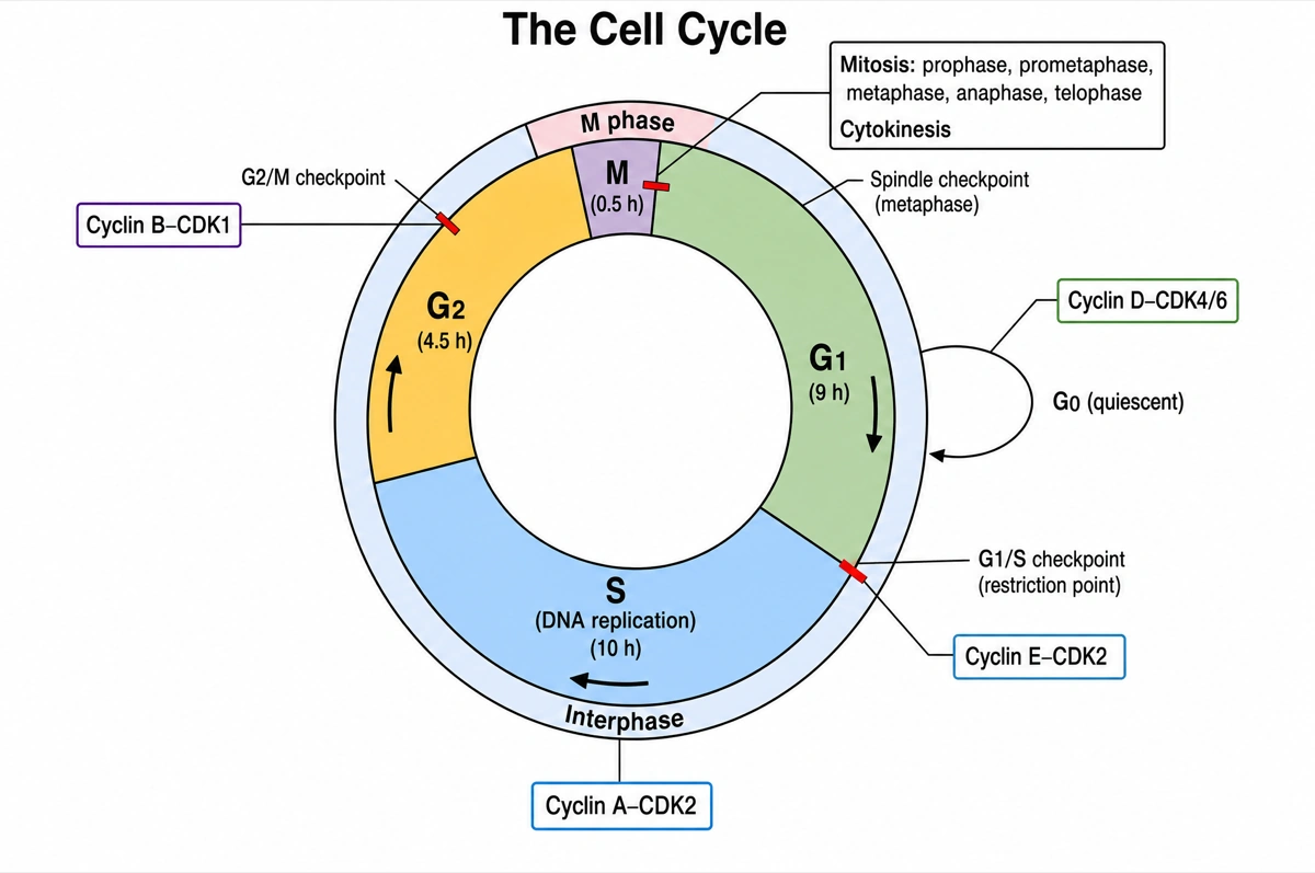
Labeled cell cycle diagram (answer key)
The eukaryotic cell cycle as one circle: interphase (G1, S, G2) and the M phase with its mitotic stages and cytokinesis, G0 to the side, the three checkpoints and the cyclin–CDK pairs. Use it as the answer key or as a cell cycle template.
Blank cell cycle diagram to label
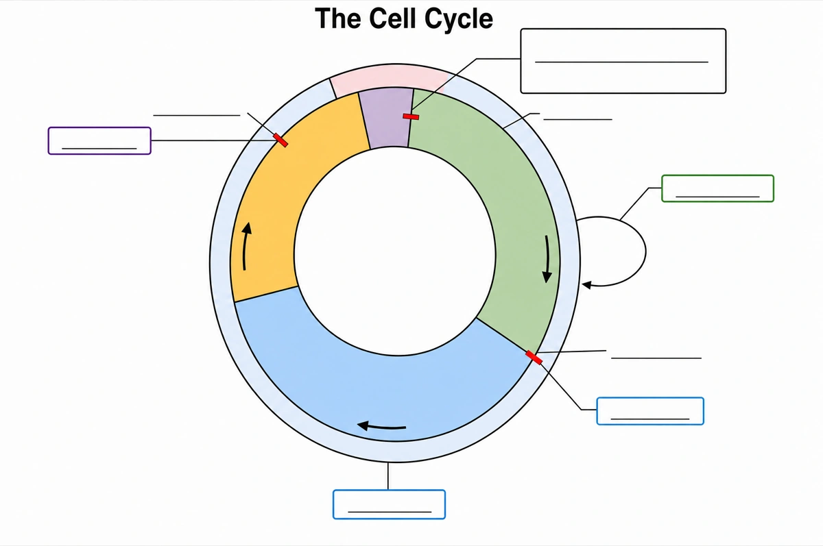
The same circle with an empty answer line in place of every phase, checkpoint and regulator name, ready for a worksheet: students name each phase, then mark where the cell checks itself before going on.
What is the cell cycle?
The cell cycle is the ordered series of events in which a eukaryotic cell grows, copies its DNA and divides into two identical daughter cells: interphase — G1, S and G2 — followed by the mitotic (M) phase of mitosis and cytokinesis, with checkpoints at G1/S, G2/M and metaphase that halt the cycle until conditions are right. Cyclin–CDK complexes drive the cell from one phase to the next.
Key facts
- Interphase takes almost all of the time. In rapidly dividing human cells with a 24-hour cycle, G1 lasts about 9 hours, S about 10 hours, G2 about 4.5 hours and M only about half an hour — which is why a correct diagram draws M as a thin slice.
- DNA is copied once, in S phase. Each chromosome is replicated into two sister chromatids joined at the centromere, and the centrosome is duplicated too; G1 and G2 are growth and preparation gaps on either side.
- Three checkpoints guard the cycle. The G1 checkpoint (restriction point) checks size, reserves, growth signals and DNA damage; the G2/M checkpoint checks that the DNA is fully replicated and undamaged; the spindle checkpoint holds anaphase until every kinetochore is attached to spindle fibers from opposite poles.
- Cyclins rise and fall; CDKs stay. CDK levels stay fairly constant while cyclins come and go: cyclin D–CDK4/6 in early G1, cyclin E–CDK2 at the G1/S transition, cyclin A–CDK2 during S phase and cyclin B–CDK1 at the G2/M transition.
- Cells can leave the cycle into G0. Quiescent cells wait in G0 until a signal returns them to G1; cells that never or rarely divide, such as mature cardiac muscle and nerve cells, stay in G0 permanently.
How does the cell cycle diagram maker work?
Describe the cycle
Say what you need — the full labeled cycle, a simple interphase-and-mitosis circle, the checkpoints and cyclin–CDK pairs, or a blank worksheet. Name the phases and regulators you want and only those are drawn.
Pick a style
The default is a clean flat textbook plate: one circle read clockwise, each phase a colored arc sized to its length, checkpoints as bars across the ring and every label on a straight leader line. Or apply watercolor, ink line art and other finishes.
Refine and export
The result opens in your workspace, where you can redraw regions, edit labels on the canvas, upscale and export in high resolution.
Phases of the cell cycle
The phases and checkpoints a labeled cell cycle diagram is normally expected to show, what happens in each and what drives or checks it. This is the default content unless you ask for something else.
| Phase | Part of | What happens | Driven or checked by |
|---|---|---|---|
| G0 | Outside the cycle | Quiescent: the cell carries out its job without preparing to divide | Growth signals return it to G1 |
| G1 (first gap) | Interphase | The cell grows and gathers building blocks and energy for DNA replication | Cyclin D–CDK4/6 |
| G1/S checkpoint | End of G1 | Checks size, reserves, growth signals and DNA damage; the restriction point | p53, p21, Rb; cyclin E–CDK2 lets the cell pass |
| S (synthesis) | Interphase | DNA is replicated into sister chromatids; the centrosome is duplicated | Cyclin A–CDK2 |
| G2 (second gap) | Interphase | The cell makes the proteins needed for mitosis | Cyclin B–CDK1 builds up |
| G2/M checkpoint | End of G2 | Checks that all DNA is replicated and undamaged | Cyclin B–CDK1 triggers entry into mitosis |
| Prophase | M phase (mitosis) | Chromosomes condense; the spindle starts to form | Cyclin B–CDK1 |
| Prometaphase | M phase (mitosis) | The nuclear envelope breaks down; kinetochores attach to spindle fibers | Cyclin B–CDK1 |
| Metaphase | M phase (mitosis) | Chromosomes line up at the metaphase plate | Spindle checkpoint |
| Anaphase | M phase (mitosis) | Sister chromatids separate and move to opposite poles | APC/C, once the spindle checkpoint is satisfied |
| Telophase | M phase (mitosis) | Nuclear envelopes re-form; chromosomes decondense | — |
| Cytokinesis | M phase | The cytoplasm divides: a cleavage furrow in animal cells, a cell plate in plant cells | — |
Arcs follow the timing: in a 24-hour human cell, G1 ≈ 9 h, S ≈ 10 h, G2 ≈ 4.5 h, M ≈ 0.5 h. Timings vary between cell types, so ask for the numbers your course uses and they are drawn instead.
Cell cycle vs mitosis: what is the difference?
The cell cycle is the whole life of a dividing cell — growth, DNA replication and division — while mitosis is only the division of the nucleus inside its M phase. Mitosis takes about half an hour of a 24-hour human cell cycle; interphase takes the rest.
| Cell cycle | Mitosis | |
|---|---|---|
| What it covers | Interphase (G1, S, G2) and M phase | Nuclear division within M phase |
| DNA replication | Yes, in S phase | No; the DNA is already copied |
| Stages | G1, S, G2, M (and G0 outside) | Prophase, prometaphase, metaphase, anaphase, telophase |
| Share of a 24-hour human cycle | All 24 hours | About 30 minutes |
| Chromosomes | Mostly semi-condensed chromatin | Condensed and visible under a light microscope |
| Checkpoints | G1/S, G2/M and spindle | The spindle checkpoint, in metaphase |
| Usual diagram | A circle | A row of cells, one per stage |
Draw the cell cycle as a circle and mitosis as a sequence. Cytokinesis is usually counted in the M phase but is a separate process from mitosis.
Where are cell cycle diagrams used?
Cell cycle diagrams are used mostly in biology teaching — from school units on cell division to university cell and cancer biology — and as figures in papers on proliferation, DNA damage and cancer drugs. The detail rises with the level: four phases for a first lesson, checkpoints and cyclin–CDK pairs for an exam or a paper.
School biology
Cell division units in middle and high school, AP Biology and A-level, usually a labeled circle plus the same diagram blank for students to label.
University cell biology
Lectures and exam questions on checkpoints, cyclins and CDKs, Rb and E2F, p53 and p21, and how the cycle is switched on and off.
Cancer research
Figures showing where a mutation or a drug interferes with a checkpoint or a cyclin–CDK complex, and at which phase the cells stop.
Posters and lab reports
A clean cell cycle as the background panel for flow cytometry results, cell-synchronization experiments or a science-fair board.
Common questions about the cell cycle
How do I draw a cell cycle diagram?
Draw a circle, divide it clockwise into G1, S, G2 and M with each arc sized to how long the phase takes, bracket G1, S and G2 as interphase, branch G0 off G1 and mark the checkpoints at G1/S, G2/M and in metaphase. With this maker you describe the version you need and the circle is drawn in that order.
What are the phases of the cell cycle?
Interphase — G1, S and G2 — and the M phase, which is mitosis (prophase, prometaphase, metaphase, anaphase, telophase) followed by cytokinesis. Cells that stop dividing leave G1 for G0.
Can I get a labeled cell cycle diagram and a blank one to label?
Yes — both are on this page free: the labeled cell cycle is the answer key and the blank one has an empty answer line for every phase, checkpoint and regulator. You can also generate a new pair at any level of detail.
What are the three cell cycle checkpoints?
The G1/S checkpoint (restriction point), the G2/M checkpoint and the spindle (M) checkpoint in metaphase. They check, in turn, whether conditions and the DNA are fit for replication, whether all DNA has been copied without damage, and whether every chromosome is attached to the spindle.
Which cyclin–CDK complexes control the cell cycle?
Cyclin D–CDK4/6 in G1, cyclin E–CDK2 at the G1/S transition, cyclin A–CDK2 during S phase and cyclin B–CDK1 at the G2/M transition. The CDK levels stay steady; the cyclin levels rise and fall, so each complex is active only in its window.
What is the G0 phase?
G0 is a resting state outside the cycle, where a cell does its job without preparing to divide. Some cells return to G1 when signaled; mature cardiac muscle and nerve cells stay in G0 permanently.
How long does each phase of the cell cycle last?
In rapidly dividing human cells with a 24-hour cycle, about 9 hours in G1, 10 in S, 4.5 in G2 and 0.5 in M. The cycle ranges from about eight minutes in early fruit-fly embryos to a lifetime spent in G0 for some human cells.
What do p53 and Rb do in the cell cycle?
Both are brakes that act mainly at the G1 checkpoint. p53 halts the cycle when it detects damaged DNA and switches on p21, which blocks cyclin–CDK complexes; Rb holds the transcription factor E2F until CDKs phosphorylate it, releasing E2F to start S phase.
What happens when cell cycle checkpoints fail?
Cells divide with damaged or wrongly separated chromosomes, and unchecked division is a route to cancer. Rb, p53 and p21 were all first found faulty in cancer cells, and TP53, the gene for p53, is mutated in more than half of human tumors.
Is the cell cycle the same as mitosis?
No. Mitosis is the division of the nucleus inside the M phase; the cell cycle also includes interphase, when the cell grows and copies its DNA. The comparison table above sets them side by side.
How is this different from a generic AI image tool or a blank diagram editor?
The cycle rules are built in. Every render is instructed to put the phases in their true order around one closed loop, join them with directional arrows and spell every phase and regulator exactly. Check the result against your course before printing; the canvas lets you move, relabel or redraw any part.
Can I use the diagrams for teaching or publication?
Yes — diagrams you generate are yours to use in worksheets, slides, handouts, posters and papers. Export high-resolution raster up to 4K depending on the model.
Sources
- The Cell Cycle — Biology 2e, section 10.2 — OpenStax
- Control of the Cell Cycle — Biology 2e, section 10.3 — OpenStax
- Cell Cycle, Mitotic (R-HSA-69278) — Reactome
- G1/S Transition (R-HSA-69206) — Reactome
Keep exploring
Our other diagram tools, and the guides behind them.
Tools

Make your cell cycle diagram
From one line of description to a labeled, printable cell cycle in about a minute.
Start drawing