
DNA Replication Diagram Maker
Describe the diagram you need and get a labeled DNA replication fork — helicase, primase, DNA polymerase, Okazaki fragments and ligase in their places, every strand end marked 5′ or 3′. Or view the labeled and blank versions below, free.
Or start from an example
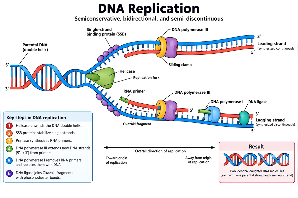
Labeled DNA replication diagram (answer key)
The replication fork with its enzymes and both new strands named. Print it as the answer key, or hand it out as a revision sheet.
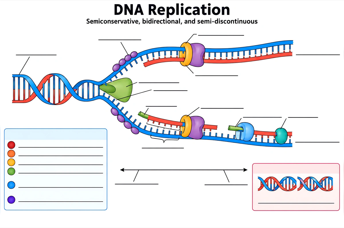
Blank DNA replication diagram to label
The same drawing with an empty answer line where every label was, ready for a worksheet, a quiz or an exam question.
What is a DNA replication diagram?
A DNA replication diagram shows how a cell copies its DNA at a replication fork: helicase separates the two strands, primase lays short RNA primers, DNA polymerase builds each new strand 5′ to 3′ — the leading strand in one piece, the lagging strand as Okazaki fragments — and DNA ligase seals the gaps. Each finished molecule keeps one old strand and one new one.
Key facts
- Replication is semiconservative. Each daughter molecule keeps one parental strand and pairs it with one newly made strand — the result Meselson and Stahl showed by growing E. coli on heavy, then light, nitrogen.
- New DNA grows only 5′ to 3′. DNA polymerase adds nucleotides only to a free 3′-OH end, so every new strand starts from a primer and runs antiparallel to its template.
- One new strand is continuous, the other is in pieces. The leading strand is built toward the fork from a single primer; the lagging strand is built away from it in short Okazaki fragments, each with its own RNA primer.
- Primers are RNA, and they are replaced. Primase makes RNA primers about 5 to 10 nucleotides long; in bacteria DNA polymerase I swaps them for DNA and DNA ligase seals the nicks that remain.
- Bacteria start from one origin, eukaryotes from many. E. coli copies its 4.6-million-base-pair chromosome from a single origin in about 42 minutes; the human genome uses up to 100,000 origins.
How does the DNA replication diagram maker work?
Describe the diagram
Say what it is for and how deep it should go — a full labeled fork, a simple overview, a close-up of the leading and lagging strands. A sentence is enough.
Pick a style
The default is a clean flat textbook plate — white background, labels on straight leader lines. Or apply watercolor, ink line art, 3D and other finishes.
Refine and export
The result opens in your workspace, where you can redraw regions, edit labels on the canvas, upscale and export in high resolution.
Enzymes of DNA replication and what each one does
The proteins a labeled DNA replication diagram is normally expected to show, what each one does, and where it sits on the fork. This is the default label set unless you ask for a shorter one.
| Enzyme or protein | What it does | Where to draw it |
|---|---|---|
| Helicase | Unwinds the double helix by breaking the hydrogen bonds between base pairs, using ATP | At the tip of the fork, where the parent helix splits |
| Single-strand binding proteins | Coat the separated strands so they do not pair up again | Along both exposed single strands |
| Topoisomerase | Relieves the over-winding ahead of the fork by nicking and resealing the DNA | On the unopened double helix ahead of the fork |
| Primase | Makes short RNA primers that give DNA polymerase a starting point | On the templates, one primer per Okazaki fragment on the lagging side |
| DNA polymerase III | Main replicating enzyme in bacteria: adds nucleotides 5′ to 3′ | On both new strands, extending from the primers |
| Sliding clamp | Ring that holds DNA polymerase on the DNA as it moves | Around the DNA at each polymerase |
| DNA polymerase I | Removes the RNA primers and fills the gaps with DNA | On the lagging strand, at the primers of finished fragments |
| DNA ligase | Seals the nicks by forming phosphodiester bonds | Between neighboring Okazaki fragments |
Courses differ in how many of these they expect. Introductory worksheets often stop at helicase, DNA polymerase and DNA ligase; advanced courses add primase, the sliding clamp and the split between DNA polymerase I and III. Name the labels you need and only those are drawn.
Leading strand vs lagging strand: what is the difference?
The leading strand is built continuously toward the replication fork from one primer; the lagging strand is built away from the fork in short Okazaki fragments, each started by its own RNA primer and later joined by DNA ligase. Both grow 5′ to 3′ — the difference comes from the two template strands running in opposite directions.
| Leading strand | Lagging strand | |
|---|---|---|
| Direction of synthesis | Toward the replication fork | Away from the replication fork |
| How it is made | Continuously, in one piece | Discontinuously, as Okazaki fragments |
| RNA primers | One, at the start | One for every Okazaki fragment |
| Template | Complementary to the 3′→5′ parental strand | Complementary to the 5′→3′ parental strand |
| Main polymerase in E. coli | DNA polymerase III | DNA polymerase III |
| Main polymerase in eukaryotes | DNA polymerase ε | DNA polymerase δ |
| After synthesis | Its single primer is replaced with DNA | Every primer is replaced and DNA ligase joins the fragments |
Draw the lagging strand as a row of short segments with arrows pointing away from the fork, the newest fragment nearest the fork still carrying its RNA primer. A lagging strand drawn as one continuous arrow, or with arrows pointing into the fork, is the error to check for first.
Where are DNA replication diagrams used?
DNA replication diagrams are used mostly in biology teaching — labeling worksheets, exam revision and lecture slides — and as schematic panels in molecular biology talks and papers. What changes between them is the depth: three labels for a first course, the full fork with the sliding clamp for a university one.
School biology
Labeling worksheets and revision sheets from middle school through GCSE, A-level, AP Biology and IB. Usually the labeled fork, plus the same drawing blank for students to fill in.
University lectures
Molecular biology and genetics slides comparing bacterial and eukaryotic replication: one origin against many, DNA polymerase III against polymerases α, δ and ε, and telomerase at the chromosome ends.
Exam revision
One-page summaries that put every enzyme at its place on the fork — which is how exam questions ask about them: name the enzyme at X, explain why the strand at Y is made in pieces.
Research and teaching figures
Schematic panels for work on replication stress, DNA repair or drugs that target topoisomerases, with the protein under study marked directly on the fork.
Common questions about DNA replication diagrams
What should a labeled DNA replication diagram include?
The standard set is the parental DNA, the replication fork, helicase, single-strand binding proteins, topoisomerase, primase and its RNA primers, DNA polymerase III, the leading strand, the lagging strand with its Okazaki fragments, DNA polymerase I and DNA ligase, with the 5′ and 3′ ends marked. That is the default — ask for fewer labels and only those are drawn.
Can I get a blank DNA replication diagram for students to label?
Yes — view the blank version on this page free, or ask for one and every label is replaced by an empty answer line. The labeled version is the matching answer key, so the two print as a pair.
Why is the lagging strand made in Okazaki fragments?
Because DNA polymerase can only build a strand 5′ to 3′, and the two template strands run in opposite directions. On one template that direction points into the fork, so synthesis is continuous; on the other it points away, so the polymerase restarts from a new primer each time the fork opens more template.
What is the difference between helicase and topoisomerase?
Helicase separates the two strands at the fork by breaking the hydrogen bonds between base pairs; topoisomerase works ahead of the fork, relieving the over-winding that the unzipping creates. It does that by making temporary nicks in the DNA and resealing them.
What does primase do, and why are RNA primers needed?
Primase makes short RNA primers, about 5 to 10 nucleotides long, that give DNA polymerase the free 3′-OH end it needs. DNA polymerase cannot start a strand on its own. The leading strand needs one primer; the lagging strand needs one for every Okazaki fragment.
What is the difference between DNA polymerase I and DNA polymerase III?
In E. coli, DNA polymerase III is the main replicating enzyme that extends both new strands; DNA polymerase I removes the RNA primers and replaces them with DNA. DNA ligase then seals the nick left between each replaced primer and the fragment beside it.
Why is DNA replication called semiconservative?
Because each new DNA molecule keeps one of the two original strands and pairs it with one newly made strand. Meselson and Stahl grew E. coli on heavy nitrogen, then light: after one generation the DNA formed a single band of intermediate density, and after two a light band appeared beside it — the pattern only semiconservative copying produces.
How is DNA replication in eukaryotes different from prokaryotes?
Eukaryotes replicate from many origins per chromosome — up to 100,000 across the human genome — at about 50 to 100 nucleotides per second, against one origin and about 1,000 nucleotides per second in E. coli. They also use other polymerases (α, δ and ε), the PCNA clamp, RNase H to remove primers, and telomerase to extend the chromosome ends.
Can I make a simple DNA replication drawing for younger students?
Yes — ask for a simple overview and the diagram drops to the essentials: the double helix unzipping, each old strand pairing with a new one, and two identical molecules at the end. Five labels or fewer usually covers a middle-school class.
How is this different from a generic AI image tool?
The subject brief is built in. Every render is instructed to follow the standard textbook drawing — both strands antiparallel, new DNA growing 5′ to 3′, Okazaki fragments only on the lagging strand — with each label spelled exactly and set on a straight leader line. Check the labels against your course before printing; the canvas lets you correct any in place.
Can I use the diagrams for school, teaching or publication?
Yes — diagrams you generate are yours to use in worksheets, presentations, handouts, theses and papers. Export high-resolution raster up to 4K depending on the model.
Can I change the labels after generating?
Yes. The result opens in a canvas workspace that recognizes the diagram labels — you can edit or translate the text, move leader lines, redraw a region or recolor without regenerating the whole figure.
Keep exploring
Our other diagram tools, and the guides behind them.
Tools


Draw your DNA replication diagram
From a one-line description to a labeled, printable diagram in about a minute.
Start drawing