
TGF-β Signaling Pathway Diagram Maker
Describe the figure you need and get a labeled TGF-β signaling pathway — active TGF-beta released from its latent complex, the type II receptor phosphorylating ALK5, SMAD2/3 joining SMAD4 in the nucleus and SMAD7 feeding back.
TGF-β signaling pathway examples
Real renders of four TGF-β signaling prompts: the fully labeled pathway with SMAD7 feedback, a six-label overview, TGF-β driving fibrosis and a blank worksheet. Click one to load its prompt and edit it.
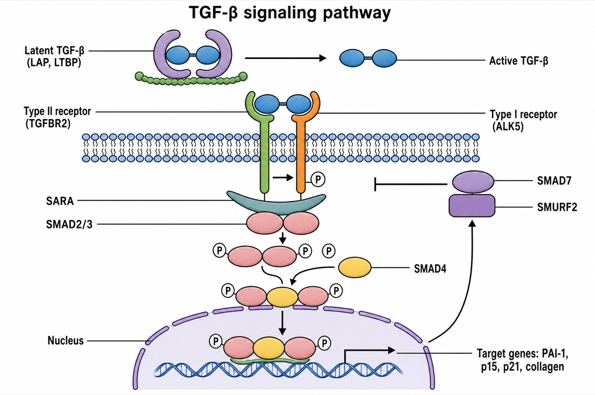
Two kinases, a SMAD complex and its own brake
Read top to bottom, then up the right side for feedback.
- 1
Latent → active TGF-β
Active TGF-β is freed from LAP and LTBP outside the cell.
- 2
TGFBR2 → ALK5
TGF-β binds TGFBR2, which phosphorylates the type I receptor ALK5.
- 3
SARA, SMAD2/3 and SMAD4
ALK5 phosphorylates SMAD2/3; they join SMAD4 and enter the nucleus.
- 4
Target genes and SMAD7
PAI-1, p15, p21, collagen switch on; SMAD7 with SMURF2 bars ALK5.
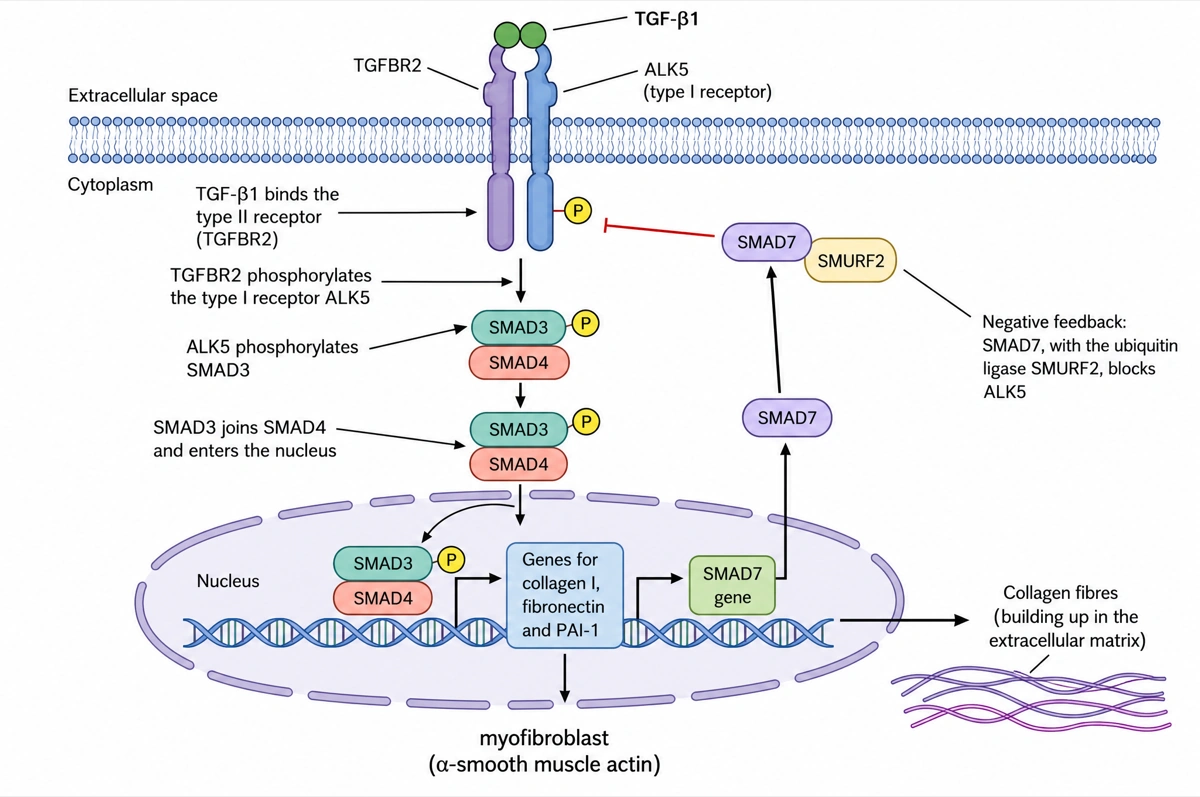
TGF-β1 drives fibrosis while SMAD7 pushes back
Read down the centre, then up the SMAD7 arm on the right.
- 1
TGFBR2 → ALK5 → SMAD3
Both receptors relay TGF-β1; SMAD3 and SMAD4 enter the nucleus.
- 2
Genes → myofibroblast
Collagen I, fibronectin, PAI-1 switch on; collagen fibres build up.
- 3
SMAD7 feedback
The SMAD7 gene is induced too; SMAD7 with SMURF2 blocks ALK5.
What is the TGF-β signaling pathway?
The TGF-β signaling pathway carries a signal from transforming growth factor beta at the cell surface to the nucleus: TGF-β binds the type II receptor (TGFBR2), which phosphorylates the type I receptor ALK5 (TGFBR1); ALK5 phosphorylates SMAD2 and SMAD3, which join SMAD4 and enter the nucleus to regulate target genes. SMAD7, one of those targets, turns the receptor back down.
Key facts
- TGF-β is made in a latent form. Furin cuts the precursor, but the latency-associated peptide (LAP) stays wrapped around the mature dimer and LTBPs anchor it in the extracellular matrix; integrins such as αvβ6 and αvβ8, proteases, thrombospondin-1 and acid release the active cytokine.
- Both receptors are serine/threonine kinases. TGF-β binds TGFBR2 first; TGFBR2 then recruits TGFBR1 (ALK5) and phosphorylates its glycine- and serine-rich GS domain, and the active complex holds two type II and two type I receptors.
- SMAD2 and SMAD3 are the R-SMADs; SMAD4 is the only co-SMAD. The adaptor SARA presents SMAD2/3 to ALK5, which phosphorylates two serines at their C-terminus; phosphorylated R-SMADs then form a complex with SMAD4 that moves into the nucleus.
- The pathway switches itself off. The SMAD2/3–SMAD4 complex induces SMAD7, which competes with SMAD2/3 for ALK5 and brings the ubiquitin ligases SMURF1, SMURF2 and NEDD4L to the receptor to have it degraded.
- The same signal suppresses early tumours and promotes late ones. In normal and pre-malignant cells TGF-β halts the cell cycle by inducing p15 and p21 and repressing MYC; in advanced carcinomas it drives epithelial–mesenchymal transition, invasion and metastasis.
How does the TGF-β signaling pathway diagram maker work?
Describe the diagram
Say what it is for and how deep it should go — the full SMAD pathway, a simple overview, the SMAD7 feedback in fibrosis. A sentence is enough.
Pick a style
The default is a clean flat textbook plate — white background, straight arrows, labels on leader lines. Or apply watercolor, ink line art, 3D and other finishes.
Refine and export
The result opens in your workspace, where you can redraw regions, edit labels on the canvas, upscale and export in high resolution.
Components of the TGF-β signaling pathway and what each one does
The molecules a labeled TGF-β pathway diagram is normally expected to show, in the order the signal passes through them. This is the default label set unless you ask for a shorter one.
| Component | What it does | Where to draw it |
|---|---|---|
| Latent TGF-β complex (LAP, LTBP) | Keeps mature TGF-β inactive and stores it in the extracellular matrix | At the top, in the extracellular space |
| Activators (integrins αvβ6 and αvβ8, proteases) | Release the active TGF-β dimer from LAP | Beside the latent complex |
| TGF-β dimer | The active ligand — TGF-β1, TGF-β2 or TGF-β3 | Above the membrane, bound to the receptors |
| Type II receptor (TGFBR2) | Kinase that binds TGF-β first and phosphorylates the type I receptor | Spanning the membrane |
| Type I receptor (ALK5, TGFBR1) | Kinase switched on by phosphorylation of its GS domain; phosphorylates SMAD2 and SMAD3 | Spanning the membrane beside TGFBR2, with a circled P |
| SARA | Adaptor that presents SMAD2/3 to ALK5 | Just under the membrane, beside ALK5 — strictly on early endosomes |
| SMAD2 and SMAD3 (R-SMADs) | Phosphorylated by ALK5 on two C-terminal serines, then released | In the cytoplasm, each with a circled P |
| SMAD4 (co-SMAD) | Shared partner that forms the SMAD2/3–SMAD4 complex | In the cytoplasm, joining the R-SMADs |
| Partner transcription factors | DNA-binding partners such as SP1 and FOXO, and co-activators such as p300, that choose the genes | On the DNA in the nucleus |
| Target genes | PAI-1 (SERPINE1), p15 (CDKN2B), p21 (CDKN1A), collagen (COL1A2), JUNB and SMAD7 on; MYC off | In the nucleus, at the bottom |
| SMAD7 with SMURF1/2 | Inhibitory SMAD that blocks ALK5 and has it degraded | A flat-headed bar from the nucleus back to ALK5 |
Courses differ in depth. An introductory figure shows TGF-β, the two receptors, SMAD2/3, SMAD4 and target genes; a cell biology course adds SARA, SMAD7 and SMURFs; advanced courses add the latent complex and the non-SMAD branches — TAK1 to p38 and JNK, PI3K to AKT. Many textbooks draw SMAD phosphorylation at the plasma membrane, while Reactome places it on early endosomes after the receptors are internalized.
TGF-β vs BMP signaling: what is the difference?
TGF-β and BMP signaling use the same machinery — a type II receptor that phosphorylates a type I receptor, R-SMADs and SMAD4 — but TGF-β signals through ALK5 and SMAD2/3, while BMPs signal through ALK2, ALK3 or ALK6 and SMAD1/5/8. Both belong to the TGF-β family, which has 33 members in humans.
| TGF-β branch | BMP branch | |
|---|---|---|
| Ligands | TGF-β1, TGF-β2, TGF-β3 (activin and Nodal use the same SMADs) | BMP2, BMP4, BMP6, BMP7, GDF5 and others |
| Type II receptor | TGFBR2 | BMPR2, ACVR2A, ACVR2B |
| Type I receptor | ALK5 (TGFBR1) | ALK3 (BMPR1A), ALK6 (BMPR1B), ALK2 (ACVR1); ALK1 for BMP9 and BMP10 |
| Binding order | Binds the type II receptor first | Can bind type I receptors alone; both types together bind far more tightly |
| R-SMADs | SMAD2, SMAD3 | SMAD1, SMAD5, SMAD8 |
| Co-SMAD | SMAD4 | SMAD4 |
| Anchoring adaptor | SARA | Endofin |
| DNA elements | Mainly the SMAD-binding element (CAGAC) | GC-rich elements as well as the SMAD-binding element |
| Extracellular control | LAP keeps TGF-β latent | Noggin and gremlin 1 trap BMPs |
| Typical roles | Growth arrest, extracellular matrix, immune regulation, fibrosis | Bone and cartilage formation; heart, kidney and blood vessel development |
Draw the two branches sharing SMAD4 and nothing else below the receptors. The classic error is SMAD1/5/8 under ALK5 or SMAD2/3 under a BMP receptor. High TGF-β concentrations can also engage ALK1 in endothelial cells, but that is an exception, not the textbook default.
Where are TGF-β signaling pathway diagrams used?
TGF-β pathway diagrams are used in cell biology and pathology teaching, and as schematic panels in fibrosis, cancer and immunology research. A first course needs the receptors and SMADs; a research figure usually adds the latent complex, the feedback loop or a drug target.
Cell biology courses
Receptor serine/threonine kinases set beside receptor tyrosine kinases: ligand, type II and type I receptors, SMADs and the nucleus, with the SMAD7 loop as the worked example of negative feedback.
Fibrosis research
Figures of fibroblasts turning into myofibroblasts under TGF-β — collagen, fibronectin, PAI-1 and α-smooth muscle actin — in lung, kidney, liver and skin fibrosis.
Cancer biology
Panels on the switch from tumour suppressor to tumour promoter: p15, p21 and MYC repression early, EMT, invasion and metastasis late, and drugs such as the ALK5 kinase inhibitor galunisertib.
Research figures and grant proposals
Schematics marking where a mutation or drug acts — TGFBR2 or SMAD4 loss, ALK5 inhibitors, integrin blockade — directly on the pathway.
Common questions about the TGF-β signaling pathway
What should a labeled TGF-β signaling pathway diagram include?
The standard set is TGF-β, the type II receptor (TGFBR2), the type I receptor ALK5 (TGFBR1) with its phosphorylation, SMAD2 and SMAD3 with circled Ps, SMAD4, the SMAD complex entering the nucleus, target genes, and SMAD7 feeding back to the receptor. Fuller figures add the latent complex (LAP, LTBP), SARA and the SMURF ubiquitin ligases.
What are the steps of the TGF-β/SMAD pathway?
Six steps: active TGF-β is released from its latent complex; it binds TGFBR2; TGFBR2 recruits and phosphorylates ALK5; ALK5 phosphorylates SMAD2 and SMAD3; phospho-SMAD2/3 bind SMAD4; and the complex enters the nucleus to regulate target genes with partner transcription factors. SMAD7, made in response, then turns the receptor down.
Is TGF-beta the same as TGF-β?
Yes — TGF-beta, TGF-β and TGF-b are three spellings of transforming growth factor beta. The name covers three human isoforms, TGF-β1, TGF-β2 and TGF-β3; pathway databases such as Reactome draw TGF-β1.
What does SMAD7 do?
SMAD7 is an inhibitory SMAD that shuts TGF-β signaling down: it binds the activated ALK5 receptor, keeps SMAD2/3 from docking, and recruits SMURF1, SMURF2 or NEDD4L to ubiquitinate the receptor for degradation. It also brings in the phosphatase PP1 to remove ALK5's phosphates. Because SMAD2/3–SMAD4 switch the SMAD7 gene on, this is a negative feedback loop.
What are the target genes of TGF-β?
Few are shared by every cell type — the feedback regulators SMAD7 and SKIL (SnoN) are the main exceptions. Well-known targets that depend on the cell include PAI-1 (SERPINE1), the CDK inhibitors p15 (CDKN2B) and p21 (CDKN1A), collagen type I (COL1A2), JUNB and SNAIL, while MYC is repressed.
Why is TGF-β both a tumour suppressor and a tumour promoter?
Because its effect depends on the cell: in normal and early tumour cells TGF-β halts the cell cycle by inducing p15 and p21 and repressing MYC, but advanced carcinomas that have lost this cytostatic arm use the remaining signal for epithelial–mesenchymal transition, invasion and metastasis. Deleting CDKN2B or weakening SMAD partners such as FOXO is one way tumours make the switch.
How does TGF-β cause fibrosis?
Excess TGF-β signaling drives fibroblasts to become myofibroblasts that make collagen, fibronectin and PAI-1 and express α-smooth muscle actin, so extracellular matrix builds up. In mice, removing SMAD3 or raising SMAD7 protects against experimental lung fibrosis.
What is non-canonical (non-SMAD) TGF-β signaling?
Non-canonical signaling is the set of SMAD-independent branches the same receptors switch on: TRAF proteins activate TAK1, which leads to p38 and JNK, and other routes activate ERK, PI3K–AKT, RHO GTPases and NF-κB. Most figures draw them as one side branch from the receptors.
How is this different from a generic AI image tool?
The subject brief is built in. Every render is instructed to lay the pathway out top to bottom — ligand, membrane receptors, SMADs, nucleus — with plain arrows for activation, flat-headed bars for inhibition and circled Ps for phosphorylation, every label spelled as in a textbook. Check the labels against your course before printing; the canvas lets you correct any in place.
Can I use the diagrams for teaching or publication?
Yes — diagrams you generate are yours to use in worksheets, presentations, handouts, theses and papers. Export high-resolution raster up to 4K depending on the model.
Can I change the labels after generating?
Yes. The result opens in a canvas workspace that recognizes the diagram labels — you can edit or translate the text, move leader lines, redraw a region or recolor without regenerating the whole figure.
Sources
- Signaling by TGF-beta Receptor Complex — pathway R-HSA-170834 — Reactome
- TGF-beta signaling pathway — KEGG PATHWAY hsa04350 — KEGG
- TGFβ signalling in context (Massagué, 2012) — Nature Reviews Molecular Cell Biology
- TGF-β signaling in health, disease and therapeutics (Deng et al., 2024) — Signal Transduction and Targeted Therapy
Keep exploring
Our other diagram tools, and the guides behind them.

Draw your TGF-β signaling pathway
From a one-line description to a labeled, printable pathway diagram in about a minute.
Start drawing