
Labeled Lymphatic System Diagram and Lymph Node Chart
Generate a labeled lymphatic system diagram of the whole body, a lymph node chart, a drainage map, a lymph node cross-section or a simple version to draw.
Lymphatic system diagram examples
Each card is a lymphatic system diagram drawn from its own prompt. They show the whole body labeled, drainage beside a flow of lymph chart, one lymph node in section and a simple lymphatic system diagram with six labels. Pick one to load its prompt.
Lymph node locations on a lymphatic system diagram, group by group
Where each group sits and what it drains. Nodes swell in the group that drains an infection, which is why a lymphatic system diagram for clinical teaching marks them by region.
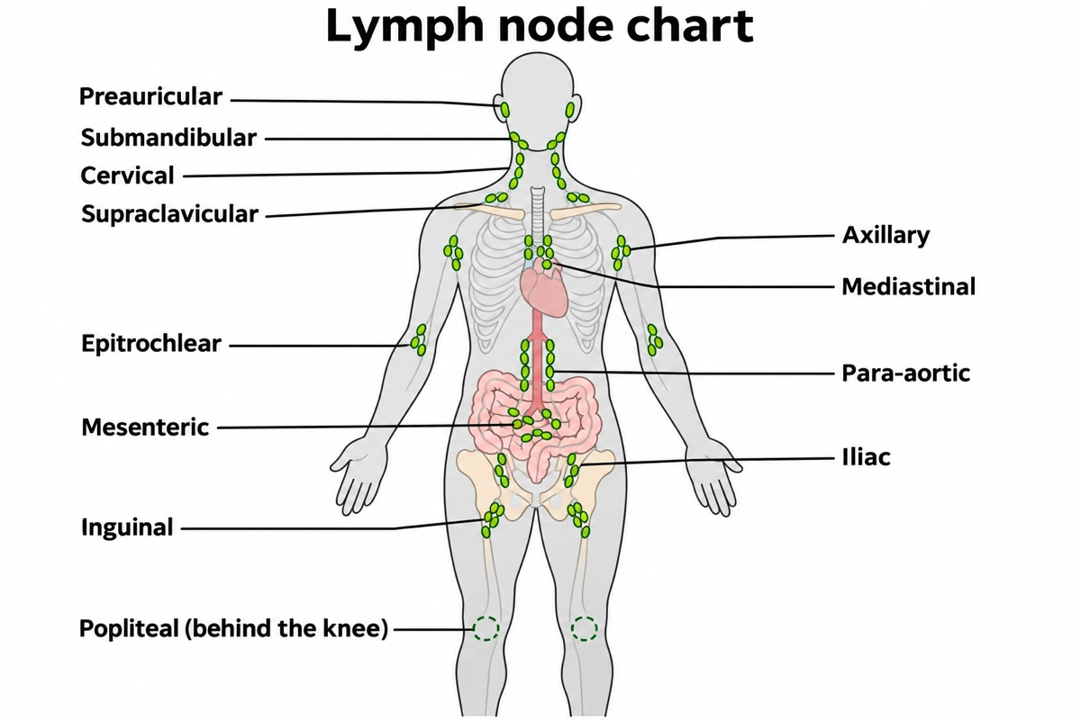
| Group on the lymphatic system diagram | Where it sits | What it drains |
|---|---|---|
| Preauricular | In front of the ear | Temple, eyelids, front of the ear, side of the face |
| Submandibular | Under the mandible | Upper lip, cheeks, body of the tongue, gums, most teeth |
| Cervical | Down the side of the neck, the deep chain on the internal jugular vein | All head and neck lymph, on its way to the jugular trunk |
| Supraclavicular | Above the collarbone | Right: chest, lungs, esophagus. Left (Virchow's node): the end of the thoracic duct, so the abdomen too |
| Axillary | Armpit, 20 to 49 nodes | Arm, lateral breast, chest wall and the abdominal wall above the navel |
| Epitrochlear | Inner arm just above the elbow | Little-finger side of the hand and forearm |
| Mediastinal | Around the trachea, bronchi and heart | Lungs, heart, esophagus, thymus |
| Para-aortic | Beside the abdominal aorta | Kidneys, testes or ovaries, back wall of the abdomen and the iliac nodes; their outflow forms the lumbar trunks |
| Mesenteric | In the mesentery | Small intestine and most of the colon, as chyle after a meal, via the intestinal trunk |
| Iliac | Along the iliac vessels in the pelvis | Bladder, rectum, uterus or prostate, and lymph already filtered by the inguinal nodes |
| Inguinal | Groin, below the inguinal ligament | Leg, buttock, abdominal wall below the navel, perineum, external genitalia; not the testes |
| Popliteal | Behind the knee, six or seven nodes | Outer foot and back of the calf along the small saphenous vein, deep tissues of the leg |
About 500 to 600 lymph nodes sit along the lymphatic vessels, most of them deep in the chest, abdomen and pelvis. Every lymphatic system diagram and textbook draws the group borders a little differently.
Lymphatic system diagrams people look for
Searches for a lymph system diagram or a diagram of the lymphatic system split into eight figures. Each entry says what a correct lymphatic system diagram of that kind shows and what to ask for.
Labeled lymphatic system diagram of the whole body
Textbooks open with a front-view lymphatic system diagram showing the vessels, the node clusters, the thoracic duct rising from the cisterna chyli to the left side of the neck, the short right lymphatic duct on the right, and the lymphoid organs. A labeled diagram of the lymphatic system for a test carries ten to fifteen names. For a lymphatic system diagram to label, ask for numbered leader lines and no names.
Lymphatic drainage map, right lymphatic duct vs thoracic duct
Drainage is lopsided. The right lymphatic duct takes only the right side of the head and neck, the right arm and the right side of the chest; the thoracic duct takes the rest. A lymphatic system diagram of drainage, the lymphatic drainage map, shades the two territories in two colours.
Flow of lymph diagram
Lymph travels from lymphatic capillaries through collecting vessels, lymph nodes, lymphatic trunks and one of the two ducts into the subclavian veins, drawn as a chart or as arrows along the vessels of a lymphatic system diagram. In a lymphatic flow diagram every arrow points toward the heart, because valves stop lymph running back.
Lymph node diagram, the structure of a lymph node
A lymph node close-up: one bean-shaped node in section, several afferent vessels entering the convex side, the subcapsular sinus, follicles with germinal centers of B cells in the cortex, T cells in the paracortex, medullary cords and sinuses, and one efferent vessel leaving at the hilum beside the artery and vein. Ask for it as an inset on a whole-body lymphatic system diagram when one slide needs both scales.
Lymph node body chart
The lymphatic system diagram used to find nodes by region: about a dozen groups on a body outline, with what each drains in the table above. A head and neck lymph nodes diagram adds the occipital, retroauricular and parotid groups.
Simple lymphatic system diagram, easy to draw
Five or six labels make a simple lymphatic system diagram: lymph nodes, lymphatic vessels, thoracic duct, tonsils, thymus and spleen. For a drawing of the lymphatic system by hand, sketch the outline, add node clusters at the neck, armpits and groin, join them with a few vessels, run the thoracic duct up the chest and put the spleen under the ribs on the person's left. An easy lymphatic system diagram leaves out the trunks and the cisterna chyli.
Primary and secondary lymphoid organs on a lymphatic system diagram
Red bone marrow and the thymus are the primary lymphoid organs, where B and T cells mature; the lymph nodes, spleen, tonsils, Peyer's patches and appendix are the secondary lymphoid organs, where those cells meet antigens. A lymphatic and immune system diagram colours the two sets differently.
Lymphatic capillaries and how lymph forms
A blind-ended lymphatic capillary among the blood capillaries, its overlapping endothelial cells working as one-way flaps. Of about 20 liters of plasma filtered from the blood each day, about 3 come back as lymph. In the small intestine these capillaries are the lacteals, which carry fat as chyle. On a full lymphatic system diagram this close-up is usually a circled inset.
What is the lymphatic system?
The lymphatic system is the network of lymphatic vessels, lymph nodes and lymphoid organs that returns excess tissue fluid to the blood, carries absorbed fat from the intestine and filters lymph for pathogens. Lymph passes some of the body's 500 to 600 lymph nodes and rejoins the blood where two ducts empty into the subclavian veins. A lymphatic system diagram draws that one-way route and the organs where lymphocytes mature.
Key facts
- B cells mature in the red bone marrow and T cells in the thymus, the two primary lymphoid organs.
- The spleen filters blood, not lymph, clearing worn-out red blood cells and blood-borne microbes.
- The thoracic duct begins at the cisterna chyli below the diaphragm, fed by the lumbar and intestinal trunks.
Right lymphatic duct vs thoracic duct on a lymphatic system diagram
The thoracic duct drains most of the body into the left subclavian vein; the right lymphatic duct drains only the upper right quarter into the right subclavian vein. This asymmetry is what a lymphatic system diagram most often gets wrong.
| Right lymphatic duct | Thoracic duct | |
|---|---|---|
| Drains | Right side of the head and neck, right arm, right side of the chest | Everything else, including the abdomen, pelvis and both legs |
| Trunks feeding it | Right jugular, right subclavian and right bronchomediastinal trunks | Lumbar and intestinal trunks through the cisterna chyli, then the left-side trunks |
| Begins | At the root of the neck | At the cisterna chyli, below the diaphragm |
| Empties into | Right subclavian vein at the right internal jugular vein | Left subclavian vein at the left internal jugular vein |
| Carries chyle | No | Yes, from the intestine |
| On a front-view lymphatic system diagram | Left side of the page | Ends on the right side of the page |
Lymphatic system diagram questions
What are the main parts of the lymphatic system?
Lymph, the lymphatic capillaries and vessels, the lymph nodes, the lymphatic trunks and two ducts, and the lymphoid organs: red bone marrow, thymus, spleen, tonsils and other lymphoid nodules. A labeled lymphatic system diagram shows all of them on one body outline.
What does the lymphatic system do?
It returns about 3 liters of leaked tissue fluid to the blood a day, carries fat absorbed in the intestine and filters lymph through the lymph nodes. On a lymphatic system diagram the vessels, lacteals and nodes stand for those three jobs.
Where are the lymph nodes located?
In clusters along the lymphatic vessels, mostly in the neck, armpits, chest, abdomen and groin. The cervical, supraclavicular, axillary and inguinal nodes can be felt when swollen; the mediastinal, para-aortic, mesenteric and iliac groups lie deep in the trunk. The lymph node chart above is a lymphatic system diagram of twelve groups and what each drains.
Where does the thoracic duct drain?
Into the left subclavian vein, where it meets the left internal jugular vein at the base of the neck. On a front-view lymphatic system diagram it ends on the right of the page.
What does the right lymphatic duct drain?
Only the right side of the head and neck, the right arm and the right side of the chest. It empties into the right subclavian vein and sits on the left of the page in a front-view lymphatic system diagram.
What is the cisterna chyli?
A sac at the foot of the thoracic duct, just below the diaphragm, that collects lymph from the legs, pelvis and abdomen through the lumbar and intestinal trunks. After a fatty meal it is milky with chyle. A lymphatic system diagram draws it as a small swelling where the thoracic duct begins.
What is lymph?
Lymph is interstitial fluid once it has entered a lymphatic capillary. It carries white blood cells, and lymph from the intestine also carries fat as milky chyle. A lymphatic system diagram often colours the lymph vessels green to set them apart from red arteries and blue veins.
How does lymph move without a heart to pump it?
Skeletal muscles squeeze the vessels, breathing changes the pressure around them, and one-way valves keep lymph moving toward the heart. That is why every arrow on a lymphatic system diagram of lymph flow points the same way, from the tissues to the subclavian veins.
What is the difference between a lymph node and the spleen?
A lymph node filters lymph; the spleen filters blood. Both are secondary lymphoid organs, but the spleen, about 12 cm long and attached to the stomach, also removes worn-out red blood cells. On a lymphatic system diagram it sits under the ribs on the person's left.
Can a lymphatic system diagram show one region only?
Yes, a lymphatic system diagram can cover one region, such as the head and neck, the armpit and breast, or the leg, with only that region's node groups and vessels labeled. Name the region, the groups and the view, front or side, in the prompt.
Sources
Keep exploring
Our other diagram tools, and the guides behind them.

Make your lymphatic system diagram
A lymphatic system diagram of the whole body, one region, the drainage map or one lymph node, labeled the way your course labels it.
Start drawing